Skin check services
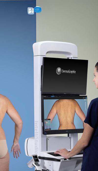
Using advanced technology to support detailed skin assessments. Discover our modern approach to skin cancer care.

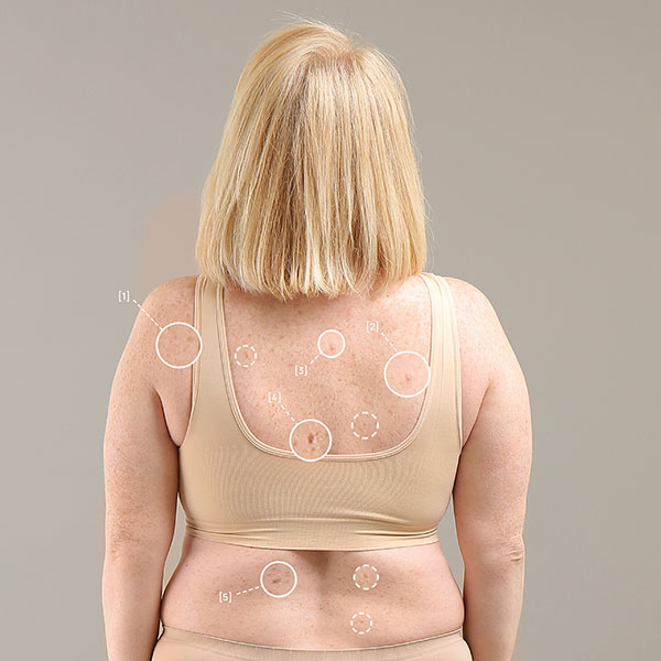
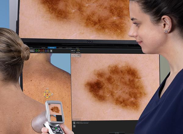
A skin check at Skintel costs $395. That includes body mapping, dermoscopy and medical review. If an optical biopsy is needed, we’ll discuss the cost and gain your consent before proceeding.
We recommend a skin check every 12 months - or sooner if you notice changes in a mole, have a personal or family history of skin cancer, or have fair skin and high sun exposure.