Skintel logo reversed

Dermoscopy

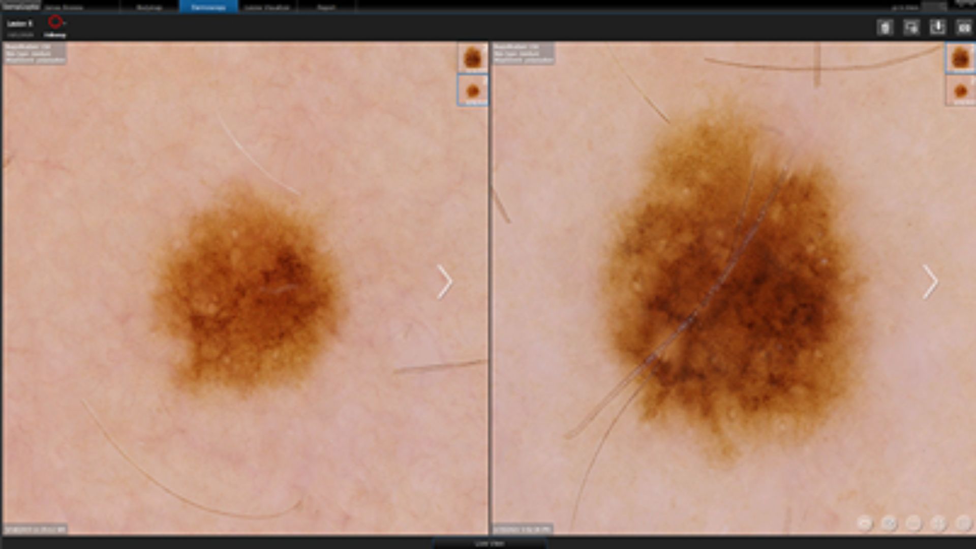
Digital dermoscopy is step 2 of your skin check, where we take a closer look at any suspicious moles at up to 200X magnification, looking through the surface of your skin to see what’s hiding below.

What is digital dermoscopy?

Dermoscopy employs powerful magnification and lighting. It enables accredited skin cancer doctors to assess the structures beneath the skin’s surface, providing critical insights that aid the detection of skin cancer.

In dermoscopy (also known as epiluminescence microscopy), we use a hand-held device to magnify your skin cells by 200x, giving us far more detail than we could see with the naked eye.

Digital dermoscopy (as used at Skintel) goes a step further, capturing high-resolution images that can be:

  • Analysed
  • Compared with past or future images of the same lesion
  • Shared with others involved in your care.

When do we use dermoscopy?

At Skintel, we use dermoscopy to investigate any suspicious moles or lesions identified during your full body scan.

Dermoscopy may be considered if your body scan detects:

  • A new or changing mole
  • A lesion with irregular borders, multiple colours or asymmetry.
  • A mole that appears inflamed, bleeding or ulcerated.

This step is crucial in skin cancer checks, as it allows for a more detailed evaluation of suspicious lesions, helping us to decide whether a biopsy is needed.

skintel-dermoscopy

How is Skintel different?

Some other skin cancer clinics rely on dermoscopy for the entirety of their skin checks, meaning you’re lying there in your underwear for a long time while a doctor checks each individual mole.

At Skintel, we see skin differently.

We begin with a full-body scan powered by artificial intelligence that maps every visible mole on your body and identifies which ones need detailed examination.

We can then use digital dermoscopy to examine specific lesions in more detail – you’re clothed by this point, which makes you feel more comfortable.

Dermoscope
© VivaScope GmbH / Oliver Gündisch

What can dermoscopy help to detect?

Melanoma

Irregular pigmentation, chaotic structures and atypical blood vessel patterns.

Basal cell carcinoma (BCC)

Ulceration, abnormal blood vessels or a pearly structure.

Squamous cell carcinoma (SCC)

Early scaling, crusting and vascular abnormalities

Non-cancerous skin conditions

Seborrheic keratosis, atypical moles, angiomas, and inflammatory skin conditions.

Next steps

After your dermoscopy, we may recommend:

Routine monitoring:

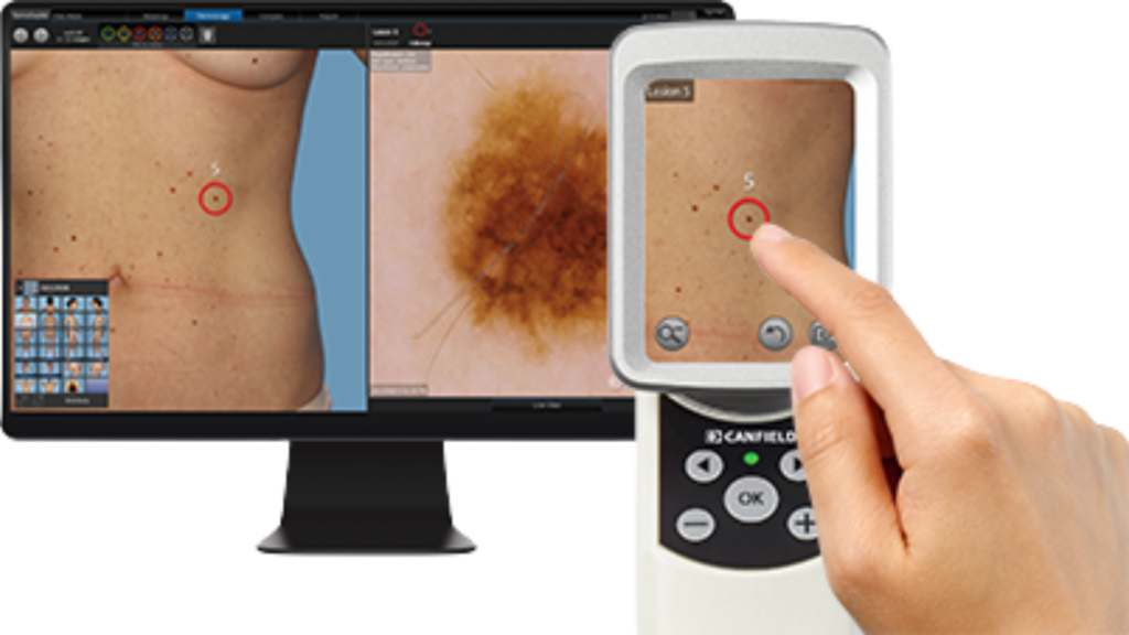
if the lesion appears low-risk, we will document it and monitor how it changes over time (e.g. compare your first body map against a future one).

Optical biopsy
More frequent skin checks:

if you have a history of skin cancer or high-risk moles, we may recommend more frequent screening.

Dermoscopy
Optical biopsy:

if a lesion raises concerns, we may conduct a non-invasive 3D optical biopsy with up to 550x magnification and same-day results.

Treatment planning:

if a lesion is highly suspicious for skin cancer, we will provide you with a detailed report to take back to your usual GP or dermatologist to consider your next steps, including surgical excision and other treatment options.

Book a skin check at Skintel

If you’ve noticed a new or changing mole or want to schedule a comprehensive skin cancer check, please book an appointment with Skintel today.

Our comprehensive skin checks include:
● A full-body map
● Digital dermoscopy (if necessary)
● Optical biopsy (if necessary).