Under the unforgiving Australian sun, it’s not unusual to notice small patches of dry, rough skin forming on areas like the face, scalp, or hands. But when these patches persist, change, or feel uncomfortable, they could be more than just sun damage. They might be actinic keratosis (AK) – a precancerous skin condition that deserves our attention.
What is actinic keratosis?
Actinic keratosis forms when long-term exposure to ultraviolet (UV) light causes abnormal changes in skin cells. These lesions typically develop in sun-exposed areas such as the face, scalp, ears, hands, and forearms. While not every lesion becomes cancerous, some can progress to squamous cell carcinoma (SCC), a common form of skin cancer.
Imagine your skin as a book that’s been left out in the sun for too long. The pages start to yellow, the cover peels, and it no longer looks as fresh as it once did. Actinic keratosis is the skin’s equivalent of those early warning signs of damage – a chance to intervene before something more serious develops.
Actinic keratosis symptoms: what to look for
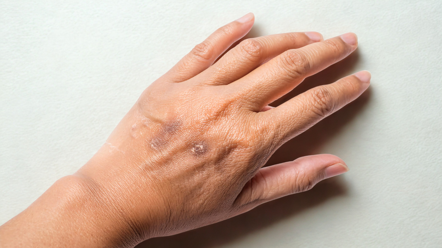
AK can present in subtle ways. What may begin as a small, flaky patch might eventually become thicker or crustier over time. You may notice:
- Rough, scaly, or crusty patches that may be pink, red, or skin-coloured
- Persistent dryness or a sandpaper-like texture that doesn’t improve with moisturiser
- Itching, burning, or tenderness in the affected area
- Lesions that change in size, texture, or appearance.
AKs can present in many different ways.
Because these symptoms often mimic dry skin or eczema, they can be easily overlooked. However, if you notice any persistent or unusual patches, it’s important to have them examined by a healthcare professional.
How is actinic keratosis diagnosed?
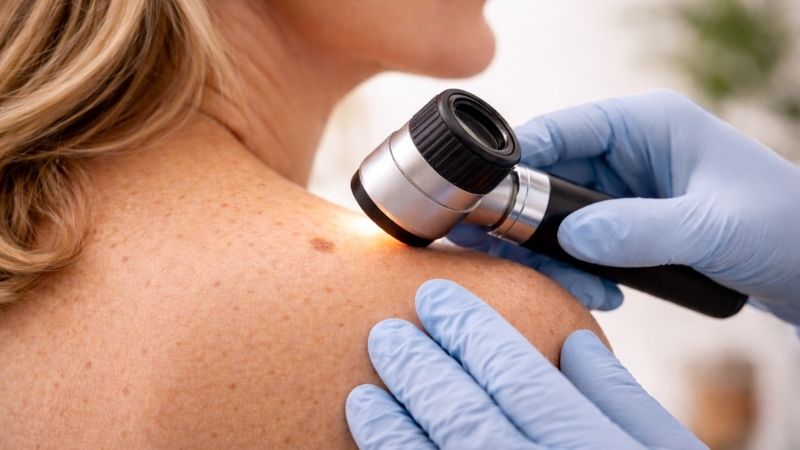
A visual skin check is often the first step. At Skintel, we use advanced diagnostic tools to examine suspicious lesions more closely.
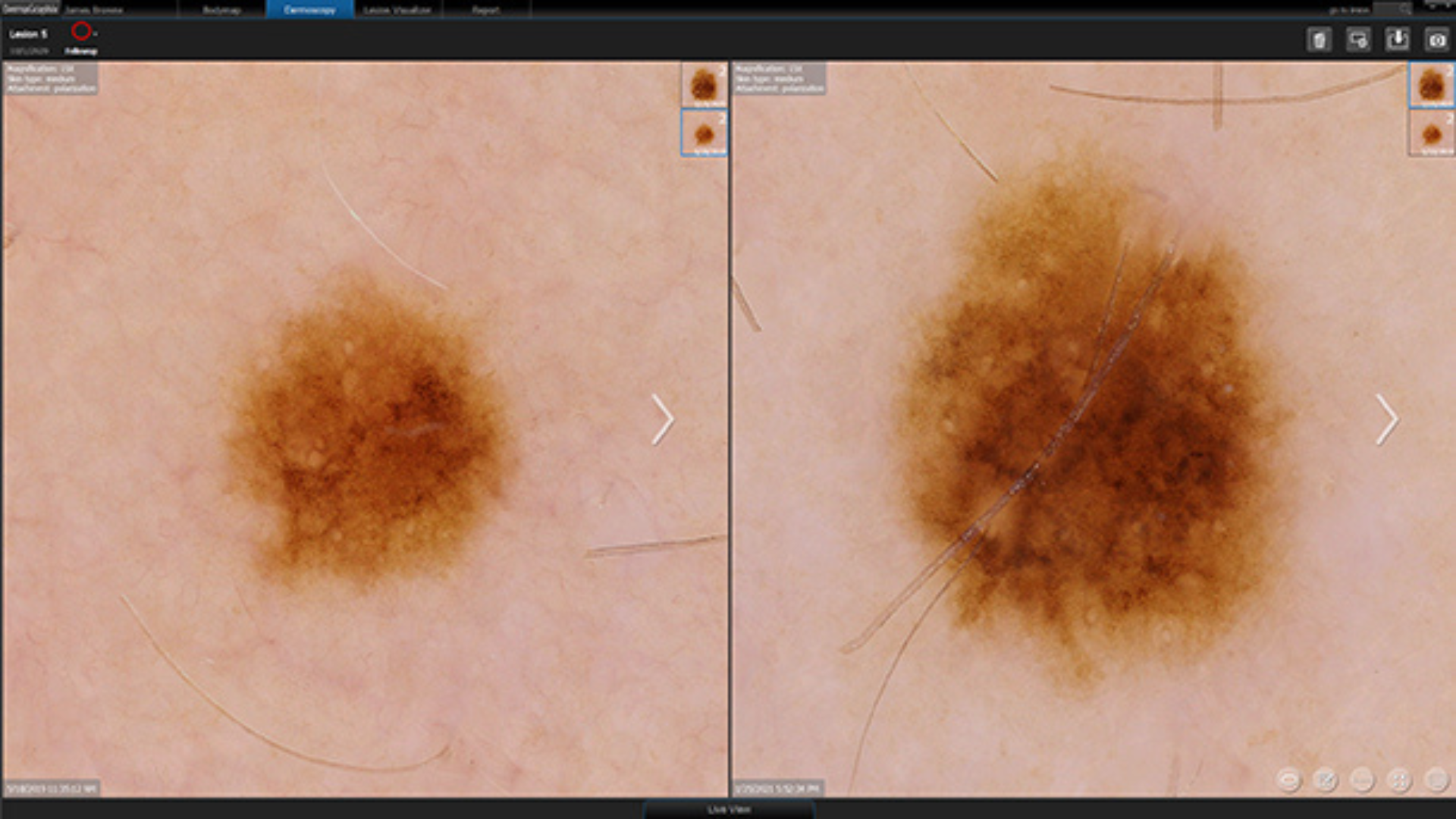
Initially, we’ll use dermoscopy to magnify and assess the structure of the skin. This is a non-invasive technique where a melanographer or accredited skin cancer doctor closely examines the lesion using a hand-held medical device. Actinic keratosis dermoscopy helps us see beneath the surface, giving us a clearer picture of whether a lesion is benign or needs further investigation.
Often, the findings are reassuring. However, if the lesion still looks suspicious, we may progress to optical biopsy. This is a non-invasive biopsy that allows us to look more deeply into your skin. At this stage, we may be trying to assess if a pre-cancerous AK has progressed to a type of skin cancer known as a squamous cell carcinoma.
See skin differently at Skintel
Actinic keratosis may be common, but it shouldn’t be ignored. Early detection is key. If you notice any unusual skin changes or would like a skin check for peace of mind, book your appointment with Skintel.
Disclaimer
All information is general and not intended as a substitute for professional advice.
References
- DermNet, Actinic keratosis, https://dermnetnz.org/topics/actinic-keratosis,[Accessed 2 July 2025]
- Healthdirect, Squamous cell carcinoma, https://www.healthdirect.gov.au/squamous-cell-carcinoma, [Accessed 2 July 2025]
- Skin Cancer College Australasia, Accreditation, https://www.skincancercollege.org/accreditation/, [Accessed 2 July 2025]