
A full-body skin cancer check is a structured examination of your skin by a trained clinician to look for melanoma, basal cell carcinoma, squamous cell carcinoma, and other concerning skin changes. It is not simply a quick look at one mole. A comprehensive check considers your skin cancer risk, examines the skin from head to toe with your consent, and may include specialised imaging such as total body photography, digital dermoscopy and, for selected lesions, reflectance confocal microscopy.¹⁻⁴
For many people, the uncertainty beforehand is the hardest part. You may wonder whether you need to undress, whether the scalp is examined, whether every mole is checked, or what happens if the clinician notices something concerning. Knowing the process in advance can make the appointment feel more straightforward.
At Skintel, a comprehensive skin check includes a technology-supported imaging pathway. Total body photography and digital dermoscopy form part of the assessment, while reflectance confocal microscopy—also called optical biopsy—is used only when it is clinically useful to clarify an uncertain lesion. A surgical biopsy is still recommended when tissue diagnosis is needed.
Before your skin cancer check
A full body skin cancer check usually begins with a conversation about your skin, your health and anything you have noticed.
The clinician may ask about:
-
new, changing, itchy, bleeding or non-healing spots;
-
previous melanoma, basal cell carcinoma or squamous cell carcinoma;
-
previous biopsies, operations or pathology results;
-
a family history of melanoma;
-
significant sun exposure, sunburns, outdoor work or tanning-bed use;
-
medications or health conditions that reduce immune function;
-
whether you have many moles or moles that have previously been described as atypical.¹⁻⁴
It is helpful to mention any specific lesion you are worried about, even if it seems minor. Bring previous pathology reports, photographs, referral letters or information about past skin cancer treatment if these are available. They can provide useful context, although you do not need to delay your appointment if you cannot find them.
How should you prepare?
There is usually no complicated preparation required for a full body skin cancer check. Wear clothing that is comfortable and easy to remove or adjust, as this makes it easier to examine the skin while preserving your privacy.
If possible, avoid heavy makeup, fake tan or thick sunscreen immediately before the appointment, particularly if you are concerned about a spot on the face. These products can make close-up examination and photography more difficult. If you have a concerning spot beneath nail polish, consider removing the polish beforehand so the nail can be assessed.
You do not need to shave body hair before a check. Hair can make some areas harder to photograph, but it is common and clinicians are used to examining hair-bearing skin. If you have noticed a lesion on the scalp, mention it during your appointment.
Make a note of any spots you want checked. Photos on your phone can be useful if a lesion has changed, bled or crusted at a time when it is not doing so during the appointment.
Do you need to undress for a full body skin check?
A thorough skin check requires access to the skin, so you will usually be asked to remove some clothing. You will be given privacy to change and can remain in underwear or use a gown or drape, depending on the clinic’s process and your preferences.
The clinician will explain what areas they plan to examine and seek your consent. You can ask questions, pause the examination or decline examination of any area.
A full body assessment may include the face, ears, neck, scalp, torso, back, arms, hands, legs, feet, soles, spaces between the toes and nails. Some melanomas and non-melanoma skin cancers occur in less visible areas, so a systematic examination is important.¹˒⁵
What happens during the full body examination?
The examination is usually organised from head to toe. The clinician looks for lesions that are new, changing, unusual compared with your other moles, or displaying features that require closer inspection.
They may use a dermatoscope, a handheld device that combines magnification with specialised polarised light. Dermoscopy allows the clinician to see pigment patterns, blood vessels and structures just below the skin surface that are not visible to the naked eye.⁶˒⁷
Not every mole needs a close-up image, but every area of skin should be assessed systematically. The clinician will focus on lesions that are visually unusual, changing, symptomatic, clinically important or different from your usual pattern of moles.
A skin check is not painful. Dermoscopy involves gentle contact with the skin or is held just above it. No needles, cutting or biopsy are involved unless a separate biopsy is later recommended by your treating doctor.
How a Skintel skin check differs
A standard skin check may involve visual examination and dermoscopy of selected lesions. At Skintel, advanced imaging is incorporated routinely into the comprehensive assessment so the skin can be reviewed at both a whole-body and close-up level. The purpose is not to replace clinical judgement, but to provide a clear visual record and additional diagnostic information for clinical review.⁸˒⁹
Total body photography and mole mapping
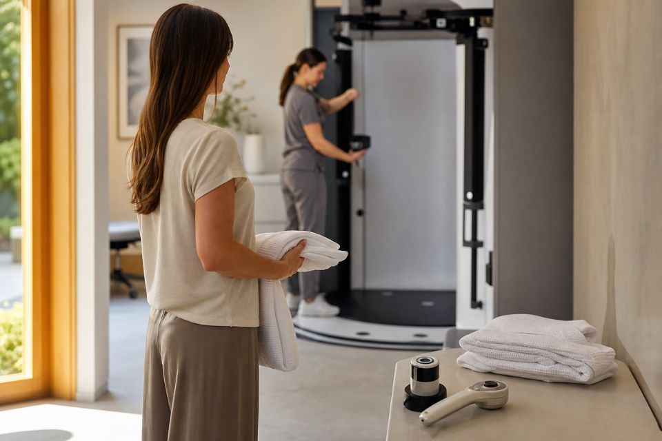
Total body photography, often called mole mapping, is a systematic set of standardised photographs of your skin. These images create a baseline record of visible moles, freckles and other skin features.¹˒²˒⁵
At future appointments, new photographs can be compared with your earlier record. This can help identify new lesions or changes in existing lesions that may otherwise be difficult to notice, particularly for people with many moles.¹˒²
Skintel uses digital mole-mapping technology to organise these photographs and link them with close-up lesion images. The software helps document and compare images, but it does not independently diagnose skin cancer. Every finding requires clinician review.
High-resolution digital dermoscopy
Digital dermoscopy captures detailed magnified images of selected moles or lesions. Dermoscopy uses specialised light and magnification to reveal skin structures that are not readily visible to the naked eye.⁶˒⁷
At Skintel, clinically relevant lesions can be photographed at high resolution and linked to your mole-mapping record. This helps document where the lesion is located and allows its appearance to be compared at later reviews.
Digital dermoscopy is particularly useful when a lesion needs closer assessment or planned surveillance. It does not mean that every photographed lesion is suspicious. Often, a detailed image simply provides a reliable baseline for future comparison.
Reflectance confocal microscopy and optical biopsy
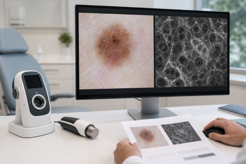
Where a lesion remains uncertain after clinical examination and dermoscopy, Skintel may use reflectance confocal microscopy, or RCM, as an additional diagnostic investigation. RCM is sometimes called an optical biopsy because it produces highly magnified black-and-white images of structures within living skin without removing tissue.¹⁰˒¹¹
RCM examines the upper layers of the skin at approximately cellular-level resolution. It may provide additional diagnostic information when a lesion is not clearly benign or clearly suspicious after other assessment.¹⁰˒¹¹
RCM is not required for every patient or every mole. It is used selectively, where the clinician believes the additional information is likely to be helpful. It also does not replace a necessary surgical biopsy. If tissue diagnosis is required, a biopsy remains appropriate because histopathology—laboratory examination of tissue removed during a biopsy—provides information that non-invasive imaging cannot always provide.¹⁰⁻¹²
A two-clinician review process
Skintel’s process involves more than one clinician. A melanographer—a nurse with specialised training in melanography and dermoscopy—conducts the imaging assessment, examines the skin during the imaging process and captures relevant total body and close-up images according to the Skintel workflow.⁸˒⁹
The images are then reviewed by an accredited skin cancer doctor, who interprets the imaging, considers the documented findings and prepares the clinical assessment. This means your skin and imaging are reviewed through a two-clinician process rather than relying on a single set of eyes.⁸˒⁹
This process does not guarantee that every skin cancer will be detected. Rather, it supports a systematic, documented assessment that combines imaging with medical review.
The Skintel imaging pathway
Not every patient needs every type of imaging. The appropriate pathway depends on your skin, the number and type of lesions present, your medical history and whether any lesion needs closer assessment.
A typical Skintel pathway may include:
-
Total body photography to document the overall skin surface and create a baseline record.
-
Digital dermoscopy to examine and document selected lesions in more detail.
-
Reflectance confocal microscopy only when a lesion remains uncertain, and the extra diagnostic information is clinically useful.
-
Surgical biopsy when tissue diagnosis is required.¹˒⁶˒¹⁰⁻¹²
This is a layered approach. It allows clinicians to look at the bigger picture, assess individual lesions closely and use additional imaging selectively rather than assuming one test suits every lesion.
Is mole mapping the same as a skin cancer check?
No. Mole mapping and a skin cancer check are related, but they are not exactly the same.
A skin cancer check is the clinical assessment of your skin by a trained clinician. It includes examining your skin, considering your risk factors and deciding whether any lesions need closer review, monitoring, biopsy or referral.
Mole mapping is the process of taking and organising systematic photographs of the skin, often with digital close-up images of selected lesions. Its key value is creating a baseline and enabling comparison over time.¹˒²˒⁵
At Skintel, total body photography and mole mapping are integrated into the comprehensive skin-check process. This provides both an immediate assessment and a visual record that may support future surveillance.
What happens if a suspicious lesion is found?
Finding a suspicious lesion does not automatically mean you have skin cancer. It means that the lesion needs more attention.
Depending on the appearance of the lesion and your clinical history, possible next steps may include:
-
reassurance and routine follow-up;
-
close-up digital dermoscopy documentation;
-
repeat imaging at a planned interval;
-
reflectance confocal microscopy if the lesion remains uncertain and is suitable for imaging;
-
a recommendation that your treating doctor arrange surgical biopsy;
-
referral back to your GP, dermatologist, surgeon or other treating clinician for management.⁶˒⁷˒¹⁰⁻¹²
Skintel is a diagnostic skin imaging service. It provides imaging findings and a report to the referring doctor. Your treating clinician remains responsible for discussing treatment options, arranging biopsy where needed and coordinating ongoing care.
Who should have a full body skin cancer check?
A medical skin assessment can be appropriate for anyone with a new, changing or concerning lesion. It is especially important to seek medical advice if a spot is growing, changing in colour or shape, bleeding, itching, painful, crusted or not healing.³˒⁴
People who may benefit from regular clinician-led surveillance include those with:
-
a previous melanoma;
-
a previous basal cell carcinoma or squamous cell carcinoma;
-
many moles or atypical moles;
-
a family history of melanoma;
-
fair or sun-sensitive skin;
-
substantial cumulative sun exposure or a history of severe sunburn;
-
immune suppression due to medication or a medical condition;
-
new or changing lesions.³⁻⁵
This does not mean that people without recognised risk factors should ignore changing spots. Anyone with a concerning lesion should seek medical assessment rather than waiting for a routine check.
How often should you have a skin cancer check?
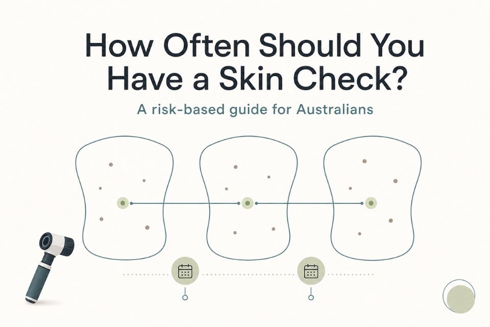
There is no single schedule that suits every Australian. Your recommended interval should be based on your personal skin cancer risk, previous findings, number of moles and your treating clinician’s advice.³˒⁴
Australian guidance commonly recommends clinician-led full body examinations every 6–12 months for people at higher risk of melanoma, supported by dermoscopy and total body photography where appropriate.¹⁻⁴ People at lower risk may need less frequent checks, while someone with a concerning new lesion should seek assessment promptly rather than waiting for a scheduled review.
Between appointments, it is sensible to remain aware of new or changing spots. If you notice something concerning, arrange an earlier review.
Safety and limitations
A comprehensive skin check is valuable, but no examination or imaging system can guarantee that every skin cancer will be detected.
Total body photography documents the skin, but does not diagnose melanoma on its own. Digital comparison tools may help identify change, but clinicians must interpret the images. Dermoscopy improves diagnostic assessment, but is not perfect. RCM can add useful information for selected uncertain lesions, but it does not replace biopsy when histopathology is required.⁶˒⁷˒¹⁰⁻¹²
The most important safety message is simple: do not wait for your next routine check if you notice a new, changing, bleeding, itchy, painful or non-healing lesion. Arrange medical assessment as soon as practical.³˒⁴
Frequently Asked Questions
What happens during a full body skin cancer check?
A full body skin cancer check begins with questions about your skin cancer history, risk factors and any spots you are concerned about. The clinician then examines the skin systematically, often using dermoscopy to inspect selected lesions more closely. At Skintel, total body photography and digital dermoscopy are integrated into the assessment, with confocal microscopy used selectively for suitable uncertain lesions.¹˒⁶˒⁸˒¹⁰
Do you have to take your clothes off for a skin check?
You will usually need to remove some clothing so the clinician can assess the skin thoroughly, but privacy and consent are always important. You can generally remain in underwear or use a gown or drape, and you should be told which areas will be examined. Intimate areas should only be examined with your clear consent and when clinically appropriate.
How long does a full body skin check take?
The length of a skin check depends on your skin type, number of moles, whether photography is performed and whether particular lesions need detailed imaging. A comprehensive imaging assessment may take longer than a brief consultation because it includes systematic photographs, close-up dermoscopic images and later medical review. Your booking confirmation should provide the most accurate appointment information for the Skintel process.
Does a skin cancer check include the scalp?
Yes, the scalp can be examined during a full body skin cancer check. The clinician may part the hair in sections or focus on areas you have noticed. Hair can make the scalp more difficult to examine and photograph, so it is useful to mention any new, scaly, tender, bleeding or changing area on the scalp.¹˒⁵
Is mole mapping the same as a skin check?
No. Mole mapping is a photographic record of the skin and selected lesions, while a skin check is a clinical examination that interprets what those images and lesions may mean. Mole mapping is particularly useful for comparing the skin over time and identifying new or changing spots.¹˒² At Skintel, mole mapping is integrated into the broader skin-check assessment.
What is total body photography?
Total body photography is a set of standardised images that records the visible skin surface. It creates a baseline record that can be compared with future photographs to help identify new lesions or changes in existing moles.¹˒²˒⁵ It does not diagnose skin cancer independently; photographs need to be reviewed with clinical examination and, where appropriate, dermoscopy.
Does every mole get checked?
Every area of skin should be assessed systematically, but not every mole needs a detailed close-up image. The clinician pays particular attention to new lesions, changing, different from your other moles, symptomatic or clinically unusual. Dermoscopy and digital photography may then be used for lesions that need closer examination.⁶˒⁷
What happens if a suspicious mole is found?
If a suspicious mole is found, the clinician may take digital dermoscopic images, recommend additional non-invasive imaging such as RCM for a suitable uncertain lesion, or advise that your treating doctor arrange a surgical biopsy.¹⁰⁻¹² A suspicious lesion does not automatically mean cancer, but it should be assessed promptly and managed according to the clinical findings.
How often should I have a skin cancer check?
The right interval depends on your individual risk. People at higher risk of melanoma are often advised to have a clinician-led full body examination every 6–12 months, with dermoscopy and total body photography where appropriate.¹⁻⁴ If you notice a new or changing lesion between scheduled appointments, seek medical advice earlier.
Conclusion
A full body skin cancer check is a structured assessment designed to look at both the overall pattern of your skin and the individual lesions that may need closer attention. It usually involves a discussion of your history, a head-to-toe examination performed respectfully and with consent, and dermoscopy of selected spots.
At Skintel, total body photography and digital dermoscopy form part of the comprehensive imaging process. Reflectance confocal microscopy may be used selectively when additional diagnostic information is needed for an uncertain lesion. When tissue diagnosis is required, surgical biopsy and histopathology remain essential.
The purpose is not to make the process complicated. It is to provide a careful, documented assessment that supports your treating clinician in making appropriate decisions about reassurance, surveillance, biopsy or referral.
Disclaimer
All information is general and not intended as a substitute for professional advice.
References
-
Menzies SW, Ingvar C, Crotty KA, McCarthy WH. Frequency and morphologic characteristics of invasive melanomas lacking specific surface microscopic features. Arch Dermatol. 1996;132(10):1178-1182.
-
Tzellos T, Kyrgidis A, Zouboulis CC. The value of total body photography for the early detection of melanoma: a systematic review. J Eur Acad Dermatol Venereol. 2021;35(2):230-237.
-
Cancer Council Australia. Early detection of skin cancer position statement. Sydney: Cancer Council Australia; 2026.
-
Royal Australian College of General Practitioners. Skin cancer. In: Guidelines for preventive activities in general practice. East Melbourne: RACGP; 2025.
-
Melanoma Institute Australia. Skin checks for melanoma position statement. Sydney: Melanoma Institute Australia; 2024.
-
Kittler H, Pehamberger H, Wolff K, Binder M. Diagnostic accuracy of dermoscopy. Lancet Oncol. 2002;3(3):159-165.
-
Menzies SW, Emery J, Staples M, et al. Impact of dermoscopy on the clinical diagnosis of melanoma in primary care: results of a randomised controlled trial. BMJ. 2009;339:b3112.
-
Skintel. The Skintel skin check. Melbourne: Skintel; 2026.
-
Skintel. About us. Melbourne: Skintel; 2026.
-
Longo C, Ragazzi M, Rajadhyaksha M, et al. Reflectance confocal microscopy: an effective tool for diagnosing melanoma and non-melanoma skin cancers. J Am Acad Dermatol. 2018;79(2):247-257.
-
Dinnes J, Bamber J, Chuchu N, et al. Reflectance confocal microscopy for diagnosing cutaneous melanoma in adults. Cochrane Database Syst Rev. 2018;12(12):CD013190. doi:10.1002/14651858.CD013190.pub2.
-
Cancer Council Australia. Clinical practice guidelines for the diagnosis and management of melanoma. Sydney: Cancer Council Australia; 2024.