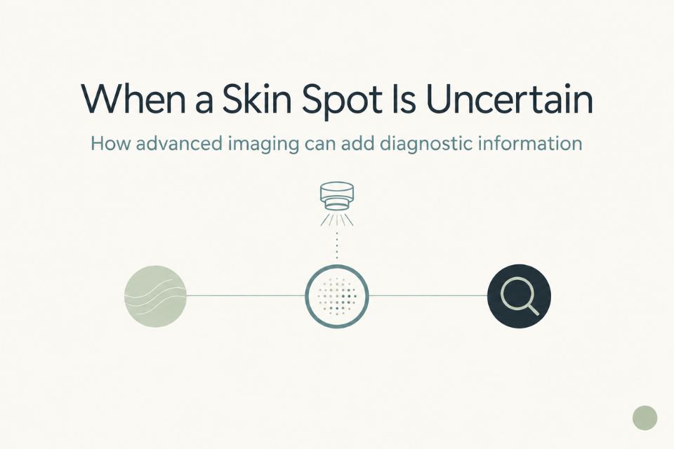

Confocal microscopy, also known as reflectance confocal microscopy (RCM), is a non‑invasive imaging test that lets your doctor see living skin cells in very fine detail without cutting the skin. A low‑power laser and a handheld microscope create “optical slices” through the top layers of your skin, producing high‑resolution, black‑and‑white images that resemble what a pathologist sees under the microscope after a biopsy.
Because the skin is not cut, RCM is often called a “virtual” or “optical” biopsy. It is used as an extra tool when dermoscopy and clinical examination alone do not give a clear answer
When is confocal microscopy used?
Confocal microscopy is not needed for every mole or spot; it is reserved for specific situations where it can meaningfully change management. Common examples include:
-
Facial or cosmetically sensitive areas – around the eyes, nose, lips or scalp, where a biopsy scar may be very noticeable.
-
Borderline or equivocal lesions – lesions that look suspicious on dermoscopy but are not clearly melanoma or clearly benign.
-
Flat facial macules and lentigo maligna – where RCM can help distinguish sun spots from early melanoma and map the true edges before surgery.
-
Monitoring non‑surgical treatment – such as topical therapies or radiotherapy for certain skin cancers.
At Skintel, confocal microscopy is integrated into a broader imaging protocol that includes full‑body imaging, dermoscopy and doctor review, rather than being used in isolation.
How accurate is confocal microscopy
When performed by trained clinicians, confocal microscopy is the most accurate non‑invasive test currently available for melanoma diagnosis. Studies from specialist centres have reported melanoma sensitivities around 92–93%, along with high accuracy for basal cell carcinoma and other skin cancers.
Crucially, RCM also helps identify many benign lesions, reducing the number of unnecessary surgical excisions. In one study, adding confocal microscopy to standard assessment reduced unnecessary biopsies by up to 43% while maintaining high melanoma detection rates.
A biopsy is still recommended whenever imaging shows concerning features or if your doctor remains unsure.
What happens during a confocal microscopy (optical biopsy) appointment
A confocal microscopy appointment is straightforward and does not involve needles, cutting or stitches. The area is cleaned, and a handheld imaging device is placed gently against your skin—often with a small amount of gel to improve contact.
The device is moved slowly to capture image “stacks” at different depths, usually over 15–30 minutes depending on how many lesions are being examined. Most people feel only light pressure from the probe, and there are no marks or scars afterwards. Confocal microscopy uses low‑power laser light and is considered safe, including for children and pregnant women in specialist centres.
Benefits and limitations
Benefits
-
Non‑invasive and scar‑sparing: no cutting, stitches or surgical wound, particularly valuable for facial lesions.
-
High diagnostic performance: very good at recognising both melanoma and common non‑melanoma skin cancers in expert hands.
-
Fewer unnecessary biopsies: helps distinguish harmless lesions from those that truly need removal.
-
Better planning: can be used to map lentigo maligna margins and support targeted biopsies or surgery.
Limitations
-
Not a full replacement for biopsy: if RCM shows suspicious changes, a traditional biopsy is still required for confirmation and to guide treatment.
-
Limited availability: currently offered only in a small number of specialist centres in Australia.
-
Best suited to certain lesions: particularly flat or slightly raised pigmented lesions; less useful for very thick, ulcerated or deeply invasive tumours.
-
Medicare: there is currently no Medicare item number for RCM in Australia, although some patients may receive support via private insurers or other schemes.
Your doctor will weigh these factors when deciding whether confocal microscopy is appropriate.
How confocal microscopy is used at Skintel in Melbourne
Skintel is one of the few dedicated skin imaging services in Australia to offer confocal laser scanning microscopy. It is used in combination with full‑body imaging and dermoscopy as part of a structured approach to skin cancer detection, rather than as a stand‑alone test.
Doctors may refer you to Skintel for confocal microscopy to:
-
Help decide whether a lesion—especially on the face—needs biopsy or can be monitored.
-
Guide the best site for biopsy within a complex lesion.
-
Support treatment planning in collaboration with your dermatologist, GP or surgeon.
Skintel focuses on diagnostic imaging and works closely with your usual treating team; confirmed skin cancers are managed by your dermatologist, surgeon or GP according to national guidelines.
Frequently asked questions
Is confocal microscopy painful?
No. Confocal microscopy is a painless test; you may only notice mild pressure from the handheld device. There are no needles, cuts or stitches, and you can return to normal activities straight away.
Can confocal microscopy replace a biopsy?
Sometimes it allows a confident diagnosis of a benign lesion, which means a biopsy can be safely avoided. If the confocal images show concerning features or your doctor is still uncertain, a traditional biopsy is still recommended to confirm the diagnosis.
Is confocal microscopy safe during pregnancy?
Confocal microscopy uses low‑power laser light and does not involve radiation or cutting the skin. It is considered safe for most people, including pregnant women and children, when performed in specialist centres.
How long does the test take?
Most confocal microscopy appointments take between 15 and 30 minutes, depending on the number and size of lesions being examined. You can usually return to normal activities immediately afterwards.
Does Medicare cover confocal microscopy?
At present, confocal microscopy is not covered by Medicare in Australia, even though it can reduce the need for unnecessary biopsies. Some patients may be able to access support through private health insurance or other schemes; your doctor or the clinic can provide more information about likely costs before you proceed
What should I do if I’m worried about a skin spot?
If you notice a new or changing spot—especially one that is growing, changing colour, itching, bleeding or looking very different from your other moles—it is important to see your GP or dermatologist promptly for a full skin examination. Confocal microscopy, if appropriate, can then be considered as part of that broader assessment.
If you or your doctor feel that a lesion is in a cosmetically sensitive area, or if there is diagnostic uncertainty, you can discuss whether referral to a skin imaging centre such as Skintel in Melbourne for confocal microscopy would be helpful.
If you would like to get in contact with us please do so here: contact us
If you would like more information about our services, please do so here: our services
References
-
Melanoma Institute Australia. Confocal Microscopy – Patient Information Leaflet.
-
Melanoma Institute Australia. Confocal Microscopy – Patient Information (online resource).
-
The Skin Hospital. Confocal Microscopy – Patient Information.
-
Sydney Melanoma Diagnostic Centre. Confocal Microscopy – Optical Biopsy Overview.
-
DermNet NZ. Reflectance confocal microscopy in dermatology.
-
Austin Clinic. Confocal Microscopy – How it works and when it is used.
-
Marghoob AA et al. Reflectance confocal microscopy made easy: The 4 must‑know key features for the diagnosis of melanoma and non‑melanoma skin cancers. Journal of the American Academy of Dermatology.
-
Soyer HP et al. Reflectance confocal microscopy in the diagnosis of nodular skin lesions. British Journal of Dermatology.
-
Melanoma Institute Australia. Deep learning for basal cell carcinoma detection in reflectance confocal microscopy (research summary).
-
Australian Government and Cancer Council melanoma and skin cancer diagnosis resources (for general statements on early detection and clinical assessment).