
By Dr Maxton Bergin
For selected skin lesions that remain uncertain after examination and dermoscopy, advanced non-invasive imaging may provide additional information before a decision is made about surgical biopsy. This can sometimes help distinguish benign lesions that may not need an invasive procedure from lesions that still require tissue sampling.
The important word is unnecessary. Imaging should never be used to delay a biopsy that is clinically indicated. When melanoma or another skin cancer is strongly suspected, or when a lesion cannot be assessed adequately with imaging, biopsy and laboratory examination of tissue remain essential.
The greatest value of advanced imaging is often in the diagnostic grey zone: lesions that are not clearly benign but are not clearly malignant after the first stages of assessment.
Can a suspicious skin spot be checked without biopsy?
Sometimes, but not always. Many skin lesions can be assessed clinically and with dermoscopy without requiring biopsy. Other lesions are clearly suspicious and should proceed to tissue diagnosis. The group in between may benefit from additional non-invasive imaging.
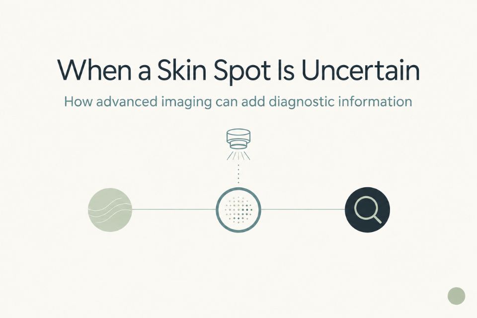
A typical pathway may look like this:
-
Clinical examination: A clinician considers the lesion’s appearance, location, symptoms, history of change and the patient’s broader skin cancer risk.
-
Dermoscopy: Magnification and specialised light reveal structures not visible to the naked eye.
-
Reflectance confocal microscopy: For selected uncertain lesions, RCM may provide additional cellular-level information without removing tissue.
-
Surgical biopsy: If tissue diagnosis is required, a clinician removes a sample or lesion for laboratory examination.
Not every lesion needs every step. The purpose is not to make the pathway longer. It is to use the appropriate level of assessment for the particular lesion.
What is dermoscopy?
Dermoscopy is a close-up examination technique that uses magnification and specialised light to examine colours, structures and patterns within a skin lesion. It can reveal details that cannot be assessed reliably with the naked eye.²
For patients, dermoscopy is usually quick and non-invasive. A handheld device may touch the skin gently or be held very close to it. It is commonly used when assessing moles, pigmented lesions, non-healing spots and lesions that may be BCC or SCC.
Dermoscopy can improve diagnostic confidence, particularly when performed by trained clinicians.² However, it cannot eliminate the need for biopsy in all cases. Some lesions remain difficult to classify even after careful dermoscopic assessment.
For a more detailed explanation, see our related article: Dermoscopy, Biopsy and Confocal: Which Test When?.
What is reflectance confocal microscopy?
Reflectance confocal microscopy, often called RCM or optical biopsy, is a non-invasive imaging technique that allows clinicians to examine structures within living skin at approximately cellular-level resolution.³
The device uses a low-power laser and does not remove tissue. There are no needles, cuts or stitches. Images are captured from the upper layers of skin and interpreted by an appropriately experienced doctor in the context of clinical examination and dermoscopy.
RCM provides additional diagnostic information. It does not universally replace histopathology. Histopathology is the laboratory examination of tissue removed during a surgical biopsy, and it remains necessary when tissue diagnosis is clinically required.
Which skin lesions may benefit from advanced imaging?
Advanced imaging is most useful when there is genuine diagnostic uncertainty. It is not required for every mole, every rough spot or every skin cancer check.
Equivocal pigmented lesions
Some moles or pigmented lesions remain uncertain after clinical examination and dermoscopy. They may have enough unusual features to prevent confident reassurance, but not enough clear signs to make immediate biopsy the only sensible option.
In these cases, RCM may help clinicians assess structures within the lesion in greater detail. Some lesions may show reassuring features on additional imaging; others may show features that support prompt biopsy.¹˒³
This is an appropriate setting for imaging-guided diagnosis because the clinical question is not “Can we avoid biopsy at all costs?” It is “Can additional information help us choose correctly between biopsy and non-invasive follow-up?”
Facial lesions and cosmetically sensitive areas
Facial lesions can be difficult because the skin is frequently sun-damaged and because an unnecessary scar can matter more on areas such as the nose, cheeks, forehead, ears and around the eyelids.
RCM may be useful for selected diagnostically uncertain facial lesions where clinical examination and dermoscopy do not provide enough confidence.⁴ It can provide additional information without immediately removing tissue.
That does not mean a suspicious facial lesion should be ignored or repeatedly imaged to avoid a scar. If biopsy is clinically required, the importance of reaching a diagnosis outweighs the disadvantages of a procedure.
For more information, see our related article: Optical Biopsy for Facial Moles and Scar-Sensitive Areas.
Lesions where basal cell carcinoma is being considered
RCM may provide useful additional information in selected lesions where basal cell carcinoma, or BCC, is part of the differential diagnosis. BCC is the most common skin cancer and can resemble benign lesions such as a scar-like patch, a pearly bump, a persistent pink area or a sun-damaged spot.⁵
A systematic review of RCM for primary BCC reported pooled sensitivity of 97% and specificity of 93%, although the authors noted limitations in the number and design of available studies.⁵ In plain language, RCM showed potential to help distinguish many BCCs from benign lookalikes in the studied settings, but it is not a universal replacement for tissue diagnosis.
Selected keratinocytic lesions
Keratinocytic lesions arise from the most common cell type in the outer layer of skin. Some sun-damaged, scaly or pink lesions may be difficult to classify, particularly when clinicians are considering benign sun damage, actinic keratosis, BCC or SCC.
Dermoscopy is usually the first imaging step. For selected lesions that remain uncertain, RCM may provide additional information, although the evidence base is stronger for some lesion types than others.³˒⁶
This is an area where clinical judgement matters. Not every rough patch needs advanced imaging, and lesions that are clearly concerning for invasive SCC may require biopsy rather than further non-invasive testing.
Benign lesions that can resemble skin cancer
Some harmless lesions can mimic skin cancer clinically or dermoscopically. Examples include certain moles, seborrhoeic keratoses, solar lentigines and inflammatory or irritated lesions.¹
This does not mean patients should try to diagnose a benign lesion themselves based on an online description. It means that a clinician may occasionally need additional information before deciding whether tissue sampling is justified.
In the right setting, RCM may improve confidence that a lesion is benign and help avoid an unnecessary procedure.¹˒³
Lesions where dermoscopy remains inconclusive
This is one of the clearest uses for advanced imaging. Dermoscopy can add considerable information, but some lesions remain indeterminate even when assessed by experienced clinicians.
In a randomised clinical trial involving 3,165 patients with dermoscopically suspicious lesions, adding RCM to standard assessment reduced the number of lesions excised per melanoma detected from 5.3 to 3.0—a 43.4% reduction in the number needed to excise.¹
“Number needed to excise” means how many lesions must be surgically removed to find one melanoma. A lower figure suggests that fewer benign lesions were removed in the process of detecting melanoma.
This is encouraging evidence, but it does not mean that RCM can rule out cancer in every situation. The study involved selected lesions, referral centres and experienced clinicians.¹
Which lesions should not have biopsy delayed?
Imaging should not delay tissue diagnosis when a lesion is strongly suspicious for melanoma or another skin cancer.
Prompt biopsy or specialist-directed management may be needed when:
-
melanoma is strongly suspected clinically or dermoscopically;
-
a lesion is rapidly changing, enlarging or becoming more irregular;
-
there is significant clinical concern despite imaging;
-
the lesion is highly raised, deeply infiltrated, markedly ulcerated or otherwise unsuitable for RCM;
-
important structures are likely to extend beyond the depth that RCM can assess; or
-
histopathology is required regardless of imaging findings.
Australian melanoma guidance states that a lesion continuing to grow or change in size, shape, colour or elevation over more than one month should be biopsied or referred for expert opinion. Suspicious raised lesions should be excised rather than monitored.⁷
Patients should not seek repeated imaging simply because they want to avoid a biopsy or scar. When biopsy is clinically necessary, it is the right next diagnostic step.
Why might avoiding an unnecessary biopsy matter?
A skin biopsy is a common and generally safe procedure. However, if a lesion is benign, avoiding an unnecessary procedure can still have worthwhile benefits.
Potential advantages include avoiding:
-
a scar;
-
local anaesthetic;
-
wound care and dressing changes;
-
inconvenience and recovery time;
-
a small risk of bleeding, infection or delayed healing; and
-
removal of tissue from a cosmetically sensitive area.
These benefits are particularly relevant on the face, ears and other visible areas. But the balance changes immediately when biopsy is clinically necessary. In that situation, the importance of reaching a definitive diagnosis outweighs the potential disadvantages of surgery.
The goal is better selection of lesions for biopsy—not fewer biopsies regardless of the clinical evidence.
What does the research say about reducing unnecessary biopsies?
The strongest evidence supports RCM as an adjunct to clinical examination and dermoscopy for selected equivocal lesions, particularly when the question is whether a lesion suspected on dermoscopy needs immediate excision.
A 2022 randomised clinical trial of 3,165 patients found that adding RCM reduced the number needed to excise by 43.4%, from 5.3 to 3.0 lesions per melanoma detected. The RCM pathway also had a higher proportion of excised lesions that proved to be melanoma.¹
A longitudinal prospective study reported that systematic second-level RCM assessment of equivocal lesions avoided excision of more than half of benign lesions in its study population.⁸
Systematic reviews and meta-analyses also suggest good diagnostic performance for RCM in selected melanocytic lesions and BCC. However, studies vary in design, lesion selection, clinician expertise and clinical setting.³˒⁵˒⁶
The most accurate patient interpretation is this: for carefully selected uncertain lesions, interpreted by experienced clinicians, RCM can provide useful information that may reduce unnecessary biopsies. It does not guarantee that a lesion is benign, and it does not replace biopsy when biopsy is needed.
How Skintel fits into the diagnostic pathway
Skintel provides advanced diagnostic skin imaging. Our skin cancer screening protocol includes full-body scanning or body mapping, dermoscopy or high-magnification assessment of selected lesions and optical biopsy using confocal microscopy where clinically appropriate. Accredited skin cancer doctors review the findings and issue a detailed report with recommendations for follow-up.⁹
For an appropriate lesion, assessment may include clinical examination, high-resolution digital dermoscopy and RCM where indicated. The imaging is performed within a workflow involving melanographers and review by accredited skin cancer doctors.⁹
Skintel is a diagnostic service. Where biopsy, excision or other treatment is clinically required, the patient returns to their referring or treating clinician for management. Imaging is used to add diagnostic information—not to delay a recommended procedure or replace necessary histopathology.
For a related explanation of diagnostic uncertainty, see our related article: Imaging for Borderline Lesions: Reducing Unnecessary Biopsies.
Could your lesion benefit from additional imaging?
If a lesion remains uncertain after initial assessment, advanced imaging may provide useful additional diagnostic information before a decision is made about biopsy. Whether it is appropriate depends on the lesion, its location, how it has changed and the clinical question being asked.
The purpose is not to promise that a procedure can be avoided. It is to help ensure that biopsy is recommended when it is needed and potentially avoid it when additional imaging is reassuring and clinically appropriate.
To arrange an assessment, Book an assessment at Skintel.
Medical safety disclaimer
This article provides general information only and is not a substitute for individual medical advice. Non-invasive imaging is not appropriate for every lesion and cannot guarantee that a lesion is benign. Biopsy remains necessary where clinically indicated. If you have a new, changing, bleeding, painful, rapidly growing or otherwise concerning lesion, seek prompt assessment from a qualified medical practitioner. Do not delay recommended biopsy or treatment simply to avoid a procedure or scar.
Frequently Asked Questions
Can imaging prevent an unnecessary skin biopsy?
For selected diagnostically uncertain lesions, imaging may provide additional information that helps a clinician decide whether biopsy is needed.¹˒³ It may therefore help avoid some unnecessary biopsies of benign lesions. It does not guarantee that a biopsy can be avoided, and it should never delay tissue diagnosis when melanoma or another skin cancer is suspected.
Can confocal microscopy replace a skin biopsy?
No. Confocal microscopy does not universally replace skin biopsy. It is a non-invasive diagnostic technique that may provide additional information for selected uncertain lesions, but biopsy remains necessary when tissue diagnosis is clinically required.³ Histopathology—the laboratory examination of removed tissue—remains the reference standard for confirming many skin cancer diagnoses.
What skin lesions can be assessed with confocal microscopy?
RCM may be helpful for selected equivocal pigmented lesions, some diagnostically uncertain facial lesions and selected lesions where BCC is being considered.¹˒⁴˒⁵ It may also add information when dermoscopy remains inconclusive. Suitability depends on the lesion’s type, depth, location, surface and degree of clinical concern. Not every lesion can be imaged adequately, and not every uncertain lesion should have biopsy deferred.
Can a facial skin cancer be diagnosed without surgery?
Sometimes, additional non-invasive imaging can provide useful diagnostic information for selected uncertain facial lesions.⁴ However, imaging is not a guaranteed alternative to biopsy. Facial lesions that are suspicious for skin cancer may still require tissue diagnosis, and the need for an accurate diagnosis outweighs the desire to avoid a scar when clinical concern is significant.
Does dermoscopy mean I will not need a biopsy?
No. Dermoscopy improves the examination of skin lesions and may increase diagnostic confidence, but it does not remove the need for biopsy in all cases.² Some lesions remain uncertain after dermoscopy, while others are sufficiently suspicious that biopsy is recommended. Dermoscopy is one part of a clinical pathway, not a guarantee that a procedure can be avoided.
What does “number needed to excise” mean?
Number needed to excise describes how many lesions are surgically removed to detect one melanoma.¹ A lower number can indicate that fewer benign lesions are being removed while still identifying melanomas. In a randomised trial of selected suspicious lesions, adding RCM reduced this number from 5.3 to 3.0.¹ This does not mean RCM is appropriate for every lesion; results depend on careful lesion selection and experienced interpretation.
Can optical biopsy show whether a mole is benign?
Optical biopsy, another term for RCM, can provide additional non-invasive information that may support a clinician’s assessment of selected moles.³ It can sometimes identify features that are reassuring or features that make biopsy more appropriate. It cannot guarantee that any lesion is benign, and a lesion may still need surgical biopsy when the clinical evidence requires tissue confirmation.
What happens if imaging is still inconclusive?
If imaging remains inconclusive, biopsy may be recommended. That is an appropriate outcome, not a failure of imaging. The aim of the assessment is to gather enough information to make the safest next decision. When uncertainty remains significant, laboratory examination of tissue through histopathology may be the most reliable way to establish a diagnosis.
Should I ask for imaging instead of a recommended biopsy?
No. If your doctor recommends biopsy because of significant concern, do not delay it simply because you hope imaging might avoid a procedure.⁷ Additional imaging may be useful for some uncertain lesions, but it is not appropriate in every situation. Discuss the reason for the recommendation with your doctor and follow the diagnostic plan that best protects your health.
Conclusion
Advanced skin imaging can be valuable for selected skin lesions in the diagnostic grey zone—those that remain uncertain after clinical examination and dermoscopy. RCM may provide additional non-invasive information that helps clinicians distinguish lesions requiring biopsy from benign lesions that may not need an invasive procedure.
This is not a promise that surgery can be avoided. The priority is always accurate diagnosis. When tissue diagnosis is clinically necessary, biopsy should proceed without delay. For the right lesion, however, imaging-guided assessment may help avoid an unnecessary biopsy while ensuring suspicious lesions receive appropriate care.
References
-
Pellacani G, Farnetani F, Ciardo S, Chester J, Kaleci S, Mazzoni L, et al. Effect of reflectance confocal microscopy for suspect lesions on diagnostic accuracy in melanoma: a randomized clinical trial. JAMA Dermatol. 2022;158(7):754-761.
-
Kittler H, Pehamberger H, Wolff K, Binder M. Diagnostic accuracy of dermoscopy. Lancet Oncol. 2002;3(3):159-165.
-
Dinnes J, Bamber J, Chuchu N, Bayliss SE, Takwoingi Y, Davenport C, et al. Reflectance confocal microscopy for diagnosing cutaneous melanoma in adults. Cochrane Database Syst Rev. 2018;12(12):CD013190.
-
Longo C, Ragazzi M, Rajadhyaksha M, Nehal K, Bennassar A, Pellacani G. Reflectance confocal microscopy: an effective tool for diagnosing melanoma and non-melanoma skin cancers. J Am Acad Dermatol. 2018;79(2):247-257.
-
Kadouch DJ, Schram ME, Leeflang MMG, Limpens J, Spuls PI, de Rie MA. In vivo confocal microscopy of basal cell carcinoma: a systematic review of diagnostic accuracy. J Eur Acad Dermatol Venereol. 2015;29(10):1890-1897.
-
Pezzini C, Kaleci S, Chester J, Pellacani G, Longo C. Reflectance confocal microscopy diagnostic accuracy for malignant melanoma in different clinical settings: systematic review and meta-analysis. J Eur Acad Dermatol Venereol. 2020;34(10):2268-2279.
-
Cancer Council Australia. Clinical practice guidelines for the diagnosis and management of melanoma. Sydney: Cancer Council Australia; 2021.
-
Pellacani G, Pepe P, Casari A, Longo C. Reflectance confocal microscopy as a second-level examination in skin oncology improves diagnostic accuracy and saves unnecessary excisions: a longitudinal prospective study. Br J Dermatol. 2014;171(5):1044-1051.
-
Skintel. The Skintel skin check. Melbourne: Skintel; accessed 2026 Sep 13.