
If you have found a changing mole, want a routine skin check or have been told you may need mole mapping, it can be difficult to know where to go. A traditional skin cancer clinic and a specialist skin imaging service may both assess skin lesions, but they often serve different—sometimes overlapping—purposes.
A skin cancer clinic commonly focuses on clinical examination, diagnosis and treatment. Many clinics can assess a suspicious lesion, perform a biopsy, remove a skin cancer or provide treatments such as cryotherapy. A specialist skin imaging service focuses more heavily on documenting the skin, using advanced imaging and providing additional diagnostic information for the referring clinician.
Neither model is automatically better. The right service depends on what you need: surveillance, advanced imaging, biopsy, treatment, ongoing management, or a combination of these.
What does a skin cancer clinic do?
A skin cancer clinic is usually a medical service that assesses skin lesions and provides management for suspected or confirmed skin cancer. Depending on the clinic, services may include:
-
clinical skin examinations;
-
dermoscopy of suspicious lesions;
-
biopsies;
-
cryotherapy for selected lesions;
-
curettage or other minor procedures;
-
surgical excision;
-
pathology arrangements;
-
treatment follow-up;
-
ongoing skin cancer surveillance.⁴˒⁵
Not every skin cancer clinic provides every service. Some clinics are led by GPs with a special interest in skin cancer medicine, while others involve dermatologists, surgeons or multidisciplinary teams. The key point is that treatment-oriented services can usually manage a lesion from assessment through to biopsy, pathology and treatment planning.
If you have a lesion that clearly needs tissue diagnosis or treatment, a skin cancer clinic, GP, dermatologist or surgeon may be the most direct pathway.
What does a skin imaging service do?
A specialist skin imaging service focuses on diagnosis, documentation and advanced non-invasive imaging. It is particularly useful when the clinical question is not simply “does this need removal?” but also “how can this lesion or this person’s skin be assessed and monitored in greater detail?”
A skin imaging service may use:
-
total body photography;
-
digital mole mapping;
-
high-resolution digital dermoscopy;
-
serial image comparison;
-
reflectance confocal microscopy, also called optical biopsy;
-
structured diagnostic reports.¹⁻³˒⁶
These tools provide additional information alongside clinical examination. They do not replace clinical judgement, and they do not eliminate the need for biopsy when tissue diagnosis is required.
For people with many moles, atypical moles, a previous melanoma or a strong family history of melanoma, photographic baseline imaging can be especially useful because it allows lesions to be compared over time.¹˒²
How Skintel differs
Skintel is a specialist diagnostic skin imaging service. Its role is to provide detailed imaging and diagnostic information to support the patient’s treating clinician, rather than to provide skin cancer treatment.
Our skin-check process combines a full-body scan, detailed examination of selected lesions and optical biopsy where an additional diagnostic imaging layer is clinically useful. The findings are reviewed by accredited skin cancer doctors, who issue a report with follow-up recommendations.⁷
Total body photography and mole mapping
Total body photography creates a baseline record of your visible skin. At future appointments, the images are compared to help identify new lesions or changes in existing lesions.¹˒²
At Skintel, full-body imaging is integrated into the skin-check process. The purpose is to map visible moles and identify lesions that may need closer examination.⁷ This does not mean software independently diagnoses melanoma. Images are reviewed by trained clinicians as part of the wider assessment.
High-resolution digital dermoscopy
Dermoscopy uses magnification and specialised light to examine structures within the upper skin layers that are not readily visible to the naked eye.⁸˒⁹ High-resolution digital dermoscopy allows selected lesions to be documented and linked to the patient’s mole-mapping record.
This can be helpful when a lesion needs detailed assessment or comparison at a future visit. Dermoscopy improves diagnostic assessment, but it is not perfect. If a lesion remains concerning or uncertain, further imaging or biopsy may be required.⁸˒⁹
Reflectance confocal microscopy and optical biopsy
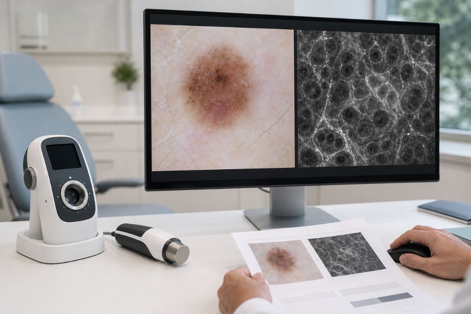
Reflectance confocal microscopy, or RCM, is a non-invasive imaging technique that produces highly magnified images of structures within living skin. It is sometimes called an optical biopsy because it allows doctors to examine cells and tissue patterns without immediately removing tissue.⁶˒¹⁰
At Skintel, RCM is used as a diagnostic investigation for selected lesions where clinical examination and dermoscopy leave meaningful uncertainty. It may provide additional diagnostic information, particularly for selected equivocal lesions and some lesions in cosmetically sensitive areas.
RCM does not replace a necessary surgical biopsy. If tissue diagnosis is clinically required, biopsy and histopathology—the laboratory examination of tissue removed during biopsy—remain essential.⁶˒¹⁰˒¹¹
Two-clinician imaging review
Our workflow involves melanographers and accredited skin cancer doctors. Melanographers are registered nurses with training in dermoscopy. They capture detailed imaging and assess the skin during the imaging process.⁷˒¹²
The images and findings are then reviewed by an accredited skin cancer doctor, who interprets the diagnostic information and provides a report with recommendations for follow-up.⁷˒¹²
This provides a documented imaging pathway with review by more than one clinician. It should not be interpreted as a guarantee that every skin cancer will be detected, but it supports systematic assessment and detailed reporting.
A diagnostic-only model
Skintel focuses on diagnostic skin imaging. If biopsy, excision, topical treatment, cryotherapy or other management is needed, the patient returns to their referring GP, dermatologist, skin cancer doctor, surgeon or other treating clinician.
This means Skintel may be part of a wider care pathway:
Skin imaging → diagnostic report → return to treating clinician → biopsy or treatment where required
The imaging service and treatment service can therefore complement each other rather than compete.
Skin cancer clinic and skin imaging service compared
| Feature | Traditional skin cancer clinic | Specialist skin imaging service |
|---|---|---|
| Full skin examination | Commonly offered | Core part of the imaging assessment |
| Dermoscopy | Commonly offered | Used with digital documentation of selected lesions |
| Total body photography / mole mapping | Available in some clinics | Central component of the Skintel imaging process |
| Serial digital comparison | Varies between clinics | Used to support comparison over time where appropriate |
| Reflectance confocal microscopy | Rarely available | Available at Skintel for selected diagnostically uncertain lesions |
| Surgical biopsy | Often available | Not performed as treatment; referring clinician manages biopsy where needed |
| Histopathology | Usually arranged after biopsy | Not performed on-site as a diagnostic test |
| Cryotherapy or other minor treatment | Often available | Not provided as part of the diagnostic imaging service |
| Excision of skin cancers | Often available | Not provided |
| Ongoing treatment follow-up | Often available | Imaging reports support the treating clinician’s plan |
| Structured diagnostic report | Varies by clinic | Provided to the referring clinician |
| Referral back to treating doctor | May not be required if treatment occurs at the clinic | Standard pathway when management is required |
Services vary between individual clinics. This table is intended as a general guide, not a statement that every clinic follows the same model.
Which service do you need?
The right service depends on the clinical question.
You may benefit from a traditional skin cancer clinic if:
-
you have a lesion that clearly requires biopsy;
-
you need treatment or excision;
-
you have already been diagnosed with skin cancer and need management;
-
you prefer assessment and treatment in the same clinic;
-
you need ongoing treatment follow-up.⁴˒⁵
You may benefit from a specialist skin imaging service if:
-
you have many moles and need systematic photographic surveillance;
-
you have atypical moles, a previous melanoma or a strong family history of melanoma;
-
a lesion is difficult to classify after examination and dermoscopy;
-
you are being referred specifically for confocal microscopy or optical biopsy;
-
you have a lesion in a cosmetically sensitive area where additional diagnostic information may be helpful before deciding whether biopsy is necessary.¹⁻³˒⁶
Sometimes you may need both
Many patients benefit from both types of service.
For example, a patient may attend Skintel for full-body imaging, mole mapping, dermoscopy and selective RCM. The diagnostic report may then recommend that a particular lesion be biopsied. The patient returns to their treating doctor, who arranges tissue diagnosis, pathology and treatment if required.
This is not duplication of care. It is a staged approach where imaging provides additional information and the treating clinician remains responsible for management.
When should a suspicious lesion go straight to biopsy?
Advanced imaging should not delay a necessary biopsy.
A lesion may need prompt biopsy when it is clinically or dermoscopically highly suspicious for melanoma or another skin cancer, when it is rapidly changing, bleeding, ulcerated, thick, nodular or otherwise unsuitable for non-invasive imaging.¹⁰˒¹¹
Histopathology remains the definitive method when tissue diagnosis is required. It can provide information about tumour type, depth and other features that help guide treatment.⁴˒⁵˒¹¹
RCM can add useful detail for selected uncertain lesions, but it is not a substitute for biopsy in every case. If the treating clinician believes tissue diagnosis is required, that decision should not be delayed simply to avoid a procedure or scar.
Is mole mapping the same as going to a skin cancer clinic?
No. Mole mapping is an imaging and surveillance tool. A skin cancer clinic is a broader clinical service that may provide skin checks, biopsies and treatment.
Some skin cancer clinics offer mole mapping, while others do not. Some specialist skin imaging services, including Skintel, integrate total body photography with digital dermoscopy and additional diagnostic imaging as part of a comprehensive assessment.¹˒²˒⁷
Mole mapping can be valuable for people at increased melanoma risk, particularly those with many moles, atypical moles or a previous melanoma. It is not a stand-alone diagnosis and should be considered alongside clinical assessment.
What happens after a skin imaging appointment?
After the imaging assessment, Skintel’s melanographers and accredited skin cancer doctors review the images and prepare a report.⁷˒¹² The report may provide reassurance, recommend routine surveillance, identify lesions for closer review or recommend that the treating doctor arrange biopsy or other management.
The report is returned to the referring clinician. Your GP, dermatologist, skin cancer doctor or surgeon then discusses the results, arranges treatment if needed and manages ongoing care.
Frequently Asked Questions
What is the difference between a skin cancer clinic and a skin imaging service?
A skin cancer clinic commonly assesses and treats skin cancers, while a specialist skin imaging service focuses more on detailed imaging, documentation and diagnostic reporting.⁴˒⁵ A skin imaging service may use total body photography, digital dermoscopy and confocal microscopy to provide additional information, then return the patient to their treating clinician for biopsy or treatment where needed.⁶˒⁷
Is mole mapping the same as a skin cancer check?
No. Mole mapping is a photographic monitoring tool that records the skin and selected lesions for comparison over time.¹˒² A skin cancer check is a clinician-led assessment that interprets lesions, risk factors and symptoms. Mole mapping can be part of a comprehensive skin check, but it does not independently diagnose melanoma.
Can a skin imaging service diagnose melanoma?
A skin imaging service can provide important diagnostic information, but a suspicious lesion may still require biopsy and histopathology for a definitive diagnosis.⁶˒¹⁰˒¹¹ Imaging supports clinical decision-making by documenting lesions and providing additional detail. It does not replace clinical judgement or tissue diagnosis when these are necessary.
Can confocal microscopy replace a biopsy?
No. Confocal microscopy can provide additional non-invasive information for selected equivocal lesions, but it cannot replace biopsy when tissue diagnosis, tumour staging or histopathology is required.⁶˒¹⁰˒¹¹ If a lesion remains suspicious or uncertain, biopsy is often the appropriate next step.
Where should I go if I have a suspicious mole?
If you have a new, changing, bleeding, itchy, painful or concerning mole, seek assessment from a GP, skin cancer doctor or dermatologist promptly.⁴˒⁵ If the lesion is clearly suspicious, direct biopsy may be appropriate. If it remains difficult to classify after clinical examination and dermoscopy, your clinician may consider referral for advanced imaging such as confocal microscopy.
Does a skin imaging clinic remove skin cancers?
A diagnostic skin imaging service such as Skintel does not remove skin cancers. Its role is to provide imaging and diagnostic reporting. If treatment is needed, the patient returns to their referring or treating clinician, who may arrange biopsy, excision, medication, cryotherapy or referral to another specialist.
Do all skin cancer clinics offer mole mapping?
No. Some skin cancer clinics provide total body photography or digital mole mapping, but availability varies.¹˒² It is worth asking whether a clinic offers baseline photography, close-up digital dermoscopy and serial image comparison if you have many moles, atypical moles, a previous melanoma or a strong family history.
What happens if imaging finds a suspicious lesion?
If imaging identifies a suspicious lesion, the next step depends on the clinical findings. The report may recommend closer assessment, confocal microscopy for an appropriate uncertain lesion, or biopsy by the treating clinician.⁶˒¹⁰˒¹¹ The treating doctor discusses the recommendation, arranges any tissue diagnosis or treatment, and coordinates follow-up.
Is mole mapping useful if I have lots of moles?
Yes. Mole mapping can be particularly useful for people with many moles because it creates a baseline record and makes it easier to identify new or changing lesions over time.¹˒² It is most valuable when interpreted by clinicians as part of regular surveillance. It does not replace assessment of any mole that changes or causes concern.
Can I use both a skin cancer clinic and a skin imaging service?
Yes. Many patients use a specialist imaging service for detailed assessment and then return to their usual doctor or skin cancer clinic for biopsy, treatment or follow-up if needed. This approach allows advanced imaging to support the management plan while keeping treatment decisions with the clinician responsible for your ongoing care.
Conclusion
A skin cancer clinic and a specialist skin imaging service can both play important roles in skin cancer care. A treatment-oriented clinic may be the best option when you need biopsy, excision or other treatment. A specialist imaging service may be particularly helpful when you need total body photography, serial monitoring, detailed digital dermoscopy or additional non-invasive assessment of an uncertain lesion.
The right choice depends on your individual needs. In many situations, the best pathway involves both: specialist imaging to provide detailed diagnostic information, followed by your treating clinician for biopsy, treatment or follow-up where required.
Disclaimer
All information is general and not intended as a substitute for professional advice.
References
-
Tzellos T, Kyrgidis A, Zouboulis CC. The value of total body photography for the early detection of melanoma: a systematic review. J Eur Acad Dermatol Venereol. 2021;35(2):230-237.
-
Ji-Xu A, Dinnes J, Matin RN. Total body photography for the diagnosis of cutaneous melanoma in adults: a systematic review and meta-analysis. Br J Dermatol. 2021;185(2):302-312.
-
Adler NR, Kelly JW, Guitera P, et al. Methods of melanoma detection and of skin monitoring for individuals at high risk of melanoma: new Australian clinical practice. Med J Aust. 2019;210(1):41-47.
-
Cancer Council Australia. Early detection of skin cancer position statement. Sydney: Cancer Council Australia; 2026.
-
Cancer Council Australia. Skin biopsy. Sydney: Cancer Council Australia; 2024.
-
Longo C, Ragazzi M, Rajadhyaksha M, et al. Reflectance confocal microscopy: an effective tool for diagnosing melanoma and non-melanoma skin cancers. J Am Acad Dermatol. 2018;79(2):247-257.
-
Skintel. The Skintel skin check. Melbourne: Skintel; 2026.
-
Kittler H, Pehamberger H, Wolff K, Binder M. Diagnostic accuracy of dermoscopy. Lancet Oncol. 2002;3(3):159-165.
-
Menzies SW, Emery J, Staples M, et al. Impact of dermoscopy on the clinical diagnosis of melanoma in primary care: results of a randomised controlled trial. BMJ. 2009;339:b3112.
-
Dinnes J, Bamber J, Chuchu N, et al. Reflectance confocal microscopy for diagnosing cutaneous melanoma in adults. Cochrane Database Syst Rev. 2018;12(12):CD013190. doi:10.1002/14651858.CD013190.pub2.
-
Cancer Council Australia. Clinical practice guidelines for the diagnosis and management of melanoma. Sydney: Cancer Council Australia; 2024.
-
Skintel. About us. Melbourne: Skintel; 2026.