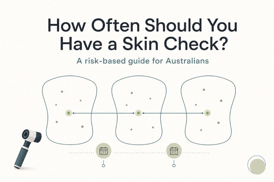

There is no single skin-check schedule that suits every adult in Australia. The right interval depends on your personal skin cancer risk, including your previous skin cancers, number and type of moles, family history, skin type, UV exposure and the findings of earlier examinations.
For people at average or lower risk, regular routine full-body skin checks are not generally recommended as a population-wide program. For people at higher risk, professional checks may be recommended every 6 to 12 months, while people at very high risk of a new melanoma may need six-monthly full-skin examination supported by total body photography and dermoscopy. Your own doctor should tailor the plan to your circumstances.¹˒²
A scheduled check is important, but it is not a reason to ignore a new or changing lesion. If a spot changes, bleeds, becomes painful, keeps itching, fails to heal or looks different from your other moles, arrange medical assessment promptly rather than waiting for your next routine appointment.
Why skin-check frequency matters in Australia
Australia has one of the world’s highest burdens of skin cancer. Ultraviolet radiation exposure is the main preventable cause, and the combination of a high-UV environment, outdoor lifestyles and individual skin characteristics means risk varies greatly from one person to another.³
Melanoma is the third most commonly diagnosed invasive cancer in Australia, while keratinocyte cancers—basal cell carcinoma and squamous cell carcinoma—are also extremely common.¹ Early assessment of suspicious lesions matters, but that does not mean every Australian needs the same screening schedule.
The useful question is not simply, “Should I get a skin check every year?” It is, “What is my risk, and what follow-up plan makes sense for me?”
Is there an official skin-check frequency in Australia?
No. Australia does not have a universal, population-wide skin cancer screening program or an official fixed interval that applies to every adult. Cancer Council Australia and the RACGP do not recommend regular skin checks for people at average or below-average risk simply because they are adults living in Australia.¹˒²
Instead, Australian guidance supports a risk-based approach:
-
People at average or lower risk should remain aware of their skin and seek assessment for any concerning new or changing lesion.
-
People at above-average risk may be offered opportunistic skin examination, generally no more often than annually unless their clinician advises otherwise.
-
People at high risk may need regular clinician-led skin checks at least every 12 months.
-
People at very high risk of new primary melanoma may need six-monthly full-skin examination supported by total body photography and dermoscopy.¹˒²
How often should high-risk patients have skin checks?
Higher-risk patients often need more regular professional surveillance, but the exact interval should be determined by their treating doctor. Australian guidance commonly supports 6–12-monthly full skin examination for people at high risk of melanoma, with six-monthly examination, total body photography and dermoscopy recommended for people at very high risk of a new primary melanoma.¹˒²
Your doctor may recommend closer review if you have a previous melanoma, multiple atypical moles, a strong family history, substantial UV damage, immune suppression or concerning findings on a recent skin examination.
Frequency is not based on one risk factor alone. It is based on the overall pattern of risk and what has been found previously.
Who may need more frequent skin checks?
People with a previous melanoma
A previous melanoma increases the risk of another melanoma as well as the risk of recurrence, so structured follow-up is important. The appropriate schedule depends on the stage and features of the original melanoma, treatments received and your risk of a second primary melanoma.⁴
For example, Australian optimal-care guidance sets out different follow-up pathways after curative treatment according to tumour thickness and stage. People with thinner melanomas may have more frequent review in the first years after treatment, while those with thicker or higher-stage melanomas may need closer specialist-directed follow-up.⁴
If you have had melanoma, follow the schedule provided by your treating team rather than relying on a general online interval.
People with previous basal cell or squamous cell carcinoma
A history of basal cell carcinoma or squamous cell carcinoma means you have already demonstrated susceptibility to UV-related skin cancer. Your clinician may recommend regular follow-up based on the number, type and location of past cancers, the extent of sun damage and whether you have ongoing risk factors such as immune suppression.¹˒³
People with many moles or atypical moles
Having many moles can make self-checking more difficult. Multiple atypical naevi—moles with unusual clinical or dermoscopic features—can also increase melanoma risk.⁵
For some people, total body photography and digital mole mapping can provide a useful baseline record. At follow-up, clinicians can compare current images with earlier images to identify new or changing lesions more objectively.⁶˒⁷
A mole count alone does not automatically determine how often you should be checked. Your clinician will consider the number of moles alongside their appearance, your personal history and your family history.
People with a strong family history
A first-degree relative—parent, sibling or child—with melanoma can increase your risk. The significance is greater if several relatives have had melanoma, if diagnoses occurred at a young age or if there is a pattern of multiple primary melanomas in the family.¹˒⁵
Tell your doctor about known family history, even if you do not have full details. It can help guide whether you need a more structured surveillance plan.
Fair skin, frequent sunburn or substantial UV exposure
Fair skin, freckling, red or fair hair, a tendency to burn, extensive outdoor work or recreation, repeated sunburn and accumulated UV exposure can all increase skin cancer risk.³
This does not mean people with darker skin cannot develop melanoma or other skin cancers. Anyone can develop skin cancer, and a concerning lesion should be assessed regardless of skin tone. Risk assessment simply helps decide whether scheduled professional surveillance should be more frequent.
People with immune suppression
People taking immune-suppressing medicines or living with conditions that reduce immune function may have a higher risk of skin cancer, particularly keratinocyte cancers. Their surveillance may need to be more structured and coordinated with the clinicians managing their broader health care.¹˒³
What about people at lower risk?
Not everyone needs a frequent professional full-body skin check. If you have few risk factors, no history of skin cancer and no concerning lesions, your doctor may recommend less frequent routine review or opportunistic examination rather than a fixed annual schedule.¹
That does not mean “do nothing”. It means using a balanced approach:
-
Be familiar with your own skin.
-
Protect your skin from UV exposure.
-
Raise new or changing lesions promptly with a doctor.
-
Review your risk if your personal or family history changes.
Routine intervals should be individualised. A person’s risk profile can change over time, particularly after a skin cancer diagnosis, significant immune suppression or the development of multiple atypical lesions.
Check your skin between appointments
Professional checks and skin imaging are valuable, but they do not replace noticing changes between visits. Skin cancers can develop between scheduled appointments, including after a recent reassuring examination.
Arrange prompt medical assessment if you notice:
-
A new mole or spot that persists.
-
A mole that has changed in size, shape, colour or elevation.
-
A lesion that looks unlike your other moles—the “ugly duckling” sign.
-
A spot that bleeds, crusts, ulcerates or does not heal.
-
Persistent itch, pain, tenderness or rapid change.
-
A dark line or changing pigment beneath a nail.³˒⁸
You do not need to diagnose the lesion yourself. The important step is noticing that something is different and arranging timely assessment.
How often should you have mole mapping?
Mole mapping does not create its own universal schedule. The frequency of total body photography, digital mole mapping and digital dermoscopy should follow your individual surveillance plan rather than a one-size-fits-all calendar.
For people at very high risk of a new melanoma, Australian guidance supports six-monthly full skin examination with total body photography and dermoscopy.¹˒² For others with many moles, atypical moles or a relevant history, imaging may be useful at an interval recommended by the clinician reviewing their overall risk.
Mole mapping can make follow-up more objective because it creates baseline images that can be compared with future images. It can help document lesion location, identify visible changes and support detailed review of selected moles.⁶˒⁷
However, mole mapping does not independently diagnose melanoma, determine your follow-up interval or replace clinical examination. A new or changing lesion still requires prompt clinician assessment.
How Skintel approaches skin surveillance
Skintel’s diagnostic workflow combines a full-body scan, detailed imaging of selected lesions and medical review. We utilise full-body scanning to map visible moles, close examination of suspicious lesions at high magnification and review by accredited skin cancer doctors, who provide a report with follow-up recommendations.⁹
Skintel’s melanographers are registered nurses trained in dermoscopy. They capture detailed skin images for assessment, while accredited skin cancer doctors review the findings and prepare the diagnostic report.¹⁰
For surveillance, total body photography and high-resolution digital dermoscopy may help create a baseline and support longitudinal comparison. The appropriate timing of repeat imaging should be tailored to the individual, based on clinical risk, previous findings and advice from the treating clinician.
If a selected lesion remains diagnostically uncertain after clinical examination and dermoscopy, reflectance confocal microscopy—also called optical biopsy—may provide additional non-invasive diagnostic information. It is not a routine part of every follow-up appointment, and it does not replace surgical biopsy when tissue diagnosis is clinically required.¹¹˒¹²
Skintel is a diagnostic skin imaging service. If biopsy, excision or other treatment is needed, management is arranged by the patient’s referring clinician.
A practical risk-based framework
The following framework is a starting point for a discussion with your doctor. It is not a substitute for an individual risk assessment.
| Risk pattern | What this may mean | Practical next step |
|---|---|---|
| Lower risk | Few recognised risk factors, no previous skin cancer and no suspicious lesion | Remain skin-aware, use sun protection and seek prompt assessment of changes. Routine professional review may be less frequent and individualised.¹ |
| Above-average risk | Several risk factors, such as fair skin, sun damage, outdoor UV exposure, family history or numerous moles | Discuss opportunistic or regular professional skin assessment with your doctor; the interval is usually tailored to the risk profile.¹ |
| High risk | Previous skin cancer, strong family history, numerous or atypical moles, substantial UV exposure or immune suppression | Regular clinician-led surveillance may be appropriate, commonly every 6–12 months depending on individual risk.¹˒² |
| Very high risk | Particularly high risk of a new melanoma, including some people with previous melanoma and extensive atypical mole patterns | Six-monthly full skin examination with total body photography and dermoscopy may be recommended.¹˒² |
No interval guarantees that a skin cancer will be detected early. The safest plan combines a clinician-recommended schedule, sensible self-awareness and prompt review when something changes.
Not sure how often you should be checked?
If you are unsure whether your skin cancer risk warrants regular surveillance, a comprehensive skin imaging assessment can help document your skin and support an individual follow-up plan with your treating clinician.
To arrange an assessment, Book a skin check at Skintel.
Medical disclaimer
This article provides general information only and is not a substitute for individual medical advice. Your personal skin-check frequency should be determined with a qualified medical practitioner who understands your history and risk factors. If you notice a new, changing, bleeding, painful or otherwise concerning lesion, arrange prompt medical assessment and do not wait for your next scheduled skin check.
Frequently Asked Questions
How often should you have a skin check in Australia?
There is no single skin-check interval for all Australians. Australia does not have a population-wide screening program or a universal recommendation that every adult needs an annual full-body skin check. People at higher risk may need professional surveillance every 6–12 months, while those at very high risk of a new melanoma may need six-monthly full skin examination with total body photography and dermoscopy.¹˒² Your doctor should tailor the interval to your risk.
Does everyone need a yearly skin check?
No. Regular annual skin checks are not generally recommended for people at average or below-average risk as a blanket population approach.¹ People with previous skin cancers, many or atypical moles, a strong family history, substantial UV exposure or immune suppression may benefit from regular professional review. If you are unsure where you fit, ask your GP or skin cancer doctor to assess your personal risk.
How often should high-risk patients have skin checks?
High-risk patients often need clinician-directed skin checks every 6–12 months, although the right interval depends on the combination of risk factors and prior findings.¹˒² People at very high risk of a new melanoma may be advised to have six-monthly full-skin examination supported by total body photography and dermoscopy. Your treating clinician may recommend a different schedule based on previous melanoma, atypical moles or other individual circumstances.
How often after melanoma should you have a skin check?
Follow-up after melanoma depends on the stage, thickness and features of the melanoma, plus your risk of another primary melanoma.⁴ Australian guidance describes closer follow-up in the early years after treatment for some stages, with timing adjusted over time. Your melanoma specialist or treating doctor should provide a written surveillance plan. Do not substitute a general online timetable for the plan designed for your diagnosis.
How often should mole mapping be repeated?
Mole mapping should be repeated according to your individual surveillance plan, not because there is one standard interval for everyone. For people at very high melanoma risk, six-monthly examination supported by total body photography and dermoscopy may be recommended.¹˒² For others, the interval may be longer and depends on mole pattern, personal history, family history and previous findings. Mole mapping supports comparison over time but does not replace clinical assessment.
Should I get checked sooner if a mole changes?
Yes. A new or changing mole should be assessed promptly rather than waiting for a routine skin check. Seek medical advice if a lesion changes in size, shape, colour or sensation; bleeds; crusts; becomes painful; does not heal; or looks different from your other moles.³˒⁸ A normal examination in the past does not rule out a new concern developing later.
Is mole mapping the same as a skin check?
No. Mole mapping is an imaging tool that documents the location and appearance of moles, often using total body photography and detailed dermoscopic images. It can help clinicians compare images over time.⁶˒⁷ A skin check is a broader clinical assessment that considers your history, skin examination, imaging where appropriate and follow-up recommendations. Mole mapping does not independently diagnose melanoma.
Can melanoma develop between annual skin checks?
Yes. Melanoma and other skin cancers can appear or change between scheduled appointments. This is why self-awareness matters even when you have regular professional checks. If a lesion is new, changing, bleeding, painful or otherwise concerning, arrange assessment promptly rather than waiting for the next annual, six-monthly or other scheduled review.³˒⁸
How often should someone with lots of moles be checked?
There is no safe universal answer based on mole number alone. Having many moles can make self-monitoring harder and may increase melanoma risk, particularly when moles are atypical or there is a personal or family history of melanoma.⁵ Your clinician may recommend regular professional surveillance and may suggest total body photography or digital dermoscopy to support image comparison over time.⁶˒⁷
Conclusion
How often you should have a skin check in Australia depends on your personal risk, not a universal annual rule. Some people need only less frequent or opportunistic review, while people at high or very high risk may require regular, clinician-directed surveillance with total body photography and dermoscopy.
The most important principle is to follow a plan tailored to your history and risk factors—and to seek prompt assessment whenever you notice a concerning change. A scheduled skin check is valuable, but it should never delay review of a new, changing, bleeding or painful lesion.
References
-
Royal Australian College of General Practitioners. Skin cancer. In: Guidelines for preventive activities in general practice. East Melbourne: RACGP; 2026.
-
Cancer Council Australia. Early detection of skin cancer position statement. Sydney: Cancer Council Australia; 2019.
-
Cancer Council Australia. Skin cancer statistics and issues: prevention policy. Sydney: Cancer Council Australia; 2026.
-
Cancer Council Australia. Optimal care pathway for people with melanoma. 2nd ed. Sydney: Cancer Council Australia; 2022.
-
Gandini S, Sera F, Cattaruzza MS, et al. Meta-analysis of risk factors for cutaneous melanoma: I. Common and atypical naevi. Eur J Cancer. 2005;41(1):28-44.
-
Ji-Xu A, Dinnes J, Matin RN. Total body photography for the diagnosis of cutaneous melanoma in adults: a systematic review and meta-analysis. Br J Dermatol. 2021;185(2):302-312.
-
Tzellos T, Kyrgidis A, Zouboulis CC. The value of total body photography for the early detection of melanoma: a systematic review. J Eur Acad Dermatol Venereol. 2021;35(2):230-237.
-
Cancer Council Australia. Checking for skin cancer. Sydney: Cancer Council Australia; 2025.
-
Skintel. The Skintel skin check. Melbourne: Skintel; 2026.
-
Skintel. About us. Melbourne: Skintel; 2026.
-
Longo C, Ragazzi M, Rajadhyaksha M, et al. Reflectance confocal microscopy: an effective tool for diagnosing melanoma and non-melanoma skin cancers. J Am Acad Dermatol. 2018;79(2):247-257.
-
Dinnes J, Bamber J, Chuchu N, et al. Reflectance confocal microscopy for diagnosing cutaneous melanoma in adults. Cochrane Database Syst Rev. 2018;12(12):CD013190.