
By Dr Maxton Bergin
An actinic keratosis is a rough, sun-damaged area of skin that can feel like sandpaper and may be easier to feel than see. It is also called a solar keratosis. Actinic keratoses are not the same as invasive skin cancer, but they are associated with long-term ultraviolet exposure and can occur in skin at increased risk of developing squamous cell carcinoma.¹˒²
A persistent rough patch, a spot that repeatedly crusts, or an area that becomes thicker, tender, ulcerated or begins bleeding should be assessed by a qualified medical practitioner. Most individual actinic keratoses do not inevitably become invasive cancer, but it is not possible to predict with certainty which lesions may progress.¹˒³
What is an actinic keratosis?
An actinic keratosis, or AK, is an area of sun-damaged skin containing abnormal keratinocytes. Keratinocytes are the main cells in the outer layer of skin. Long-term ultraviolet, or UV, exposure can damage these cells over many years, leading to rough, scaly or crusted areas of skin.¹˒²
The terms actinic keratosis and solar keratosis are commonly used for the same condition. “Actinic” means related to sunlight, while “solar” refers to the sun.
An AK is often described as a precancerous lesion or an early precursor in the pathway to cutaneous squamous cell carcinoma, known as SCC. However, this relationship needs to be understood carefully. Some AKs persist, some may regress, some recur and some may progress. Most individual lesions do not inevitably become invasive SCC.³˒⁴
Their presence is important because it tells us that the skin has accumulated significant UV damage. A person with multiple AKs may have a wider area of sun-damaged skin and a higher risk of developing additional keratinocyte cancers than someone without them.²˒³
What does actinic keratosis look and feel like?
Actinic keratosis often appears as a rough or scaly patch on sun-exposed skin. It may be pink, red, brown, skin-coloured, pale or slightly yellow-white. Some lesions are flat; others are raised or thicker.¹˒²
A useful patient description is: some actinic keratoses are noticed first by touch rather than sight. A small rough area may feel like sandpaper when you run a finger over it, even when it is difficult to see in a mirror.
An AK may also:
-
repeatedly become rough after scale is rubbed or removed;
-
feel dry, scaly or crusted;
-
remain in the same place for months;
-
be mildly tender or sensitive;
-
appear as one spot or as several patches across an area of sun-damaged skin.
Photographs and online descriptions cannot diagnose an actinic keratosis. Rough patches can have several causes, including eczema, psoriasis, harmless sun spots and different types of skin cancer. A qualified clinician needs to assess a lesion that is persistent, changing or otherwise concerning.
Where do solar keratoses occur?
Actinic keratoses usually occur on areas exposed to UV radiation over many years. Common locations include:
-
the face;
-
the scalp, particularly where hair is thinning;
-
the ears;
-
the backs of the hands;
-
the forearms;
-
the lower legs; and
-
other chronically sun-exposed sites.²˒⁵
More than 80% of AKs occur on the head, neck and upper limbs.² They can develop after years of ordinary day-to-day UV exposure, not only after obvious sunburn.
Who is most at risk?
Anyone can develop sun damage or skin cancer, but AKs are more common in people with a combination of long-term UV exposure and individual susceptibility.
Risk factors include:
-
increasing age;
-
fair or easily sunburned skin;
-
freckling;
-
substantial lifetime sun exposure;
-
outdoor work or recreation;
-
repeated UV exposure over many years;
-
a bald or thinning scalp;
-
previous actinic keratoses;
-
previous basal cell carcinoma or squamous cell carcinoma; and
-
immune suppression due to medicines or medical conditions.²˒⁵
People with darker skin can also develop actinic keratoses and skin cancer. Regardless of skin tone, a persistent, changing or concerning lesion should be assessed.
Is actinic keratosis skin cancer?
No. An actinic keratosis is not the same as invasive skin cancer. It is a sun-damaged lesion with abnormal cells in the outer layer of skin that may, in some cases, progress to invasive squamous cell carcinoma.¹˒³
A useful way to think about the relationship is:
Cumulative sun damage → actinic keratosis or solar keratosis → possible progression in some lesions → invasive squamous cell carcinoma.
This is a risk pathway, not a guaranteed sequence. Most individual AKs do not progress to invasive SCC, and it is difficult to identify in advance exactly which lesion will progress. Reported progression estimates vary widely because studies differ in the patients observed, the definition of AK and the length of follow-up.³˒⁴
The broader message is more important than a percentage: an AK indicates sun-damaged skin, and multiple AKs can be a marker of increased future risk of keratinocyte cancers.²˒³
Can actinic keratosis turn into squamous cell carcinoma?
Yes, some actinic keratoses can progress to cutaneous squamous cell carcinoma, but most individual lesions do not inevitably do so.¹˒³
It is not possible to predict reliably which individual AK will become an invasive SCC. This is why ongoing skin awareness, appropriate treatment planning with a treating clinician and reassessment of changing lesions are important.³˒⁴
Features that may justify closer assessment include:
-
rapid growth;
-
increasing thickness;
-
marked firmness or induration;
-
significant tenderness or pain;
-
persistent ulceration;
-
spontaneous bleeding;
-
a prominent horn-like keratin growth; or
-
a lesion that does not behave as expected.¹˒⁴
These features do not automatically mean that a lesion is SCC. They do mean the lesion may need closer examination and, where clinically indicated, biopsy.
Actinic keratosis vs squamous cell carcinoma
Patients cannot always distinguish an AK from SCC by looking at a lesion themselves. Both can appear as rough, scaly, crusted or red areas on sun-exposed skin.²˒⁴
An AK is generally a more superficial area of UV-damaged skin. SCC is an invasive skin cancer that can extend deeper into the skin. The difference is not always clear from a photograph or a single visual check.
Clinicians may use:
-
the lesion’s appearance and texture;
-
its location and duration;
-
whether it has changed;
-
dermoscopy;
-
the person’s history of sun damage and skin cancer; and
-
biopsy when the diagnosis is uncertain or SCC is suspected.¹˒⁴
A biopsy removes a small tissue sample so it can be examined under a microscope in a pathology laboratory. Not every AK needs biopsy, but biopsy may be needed when a lesion has concerning features or does not behave as expected.
What does “field change” mean?
Field change, also called field cancerisation, means that an entire area of chronically sun-damaged skin may contain multiple visible and microscopic abnormal areas rather than one isolated spot.²˒³
For example, someone may have several rough patches across the forehead, scalp, ears or backs of the hands. This can reflect broader cumulative UV damage in that part of the skin.
Field change helps explain why a person may develop several AKs over time and why a clinician may assess the surrounding skin—not only the one lesion that first caught the patient’s attention.
How is actinic keratosis diagnosed?
Actinic keratosis is often diagnosed through clinical examination. A clinician considers the lesion’s appearance, texture, location, duration and the person’s history of UV exposure and previous skin cancers.²˒⁴
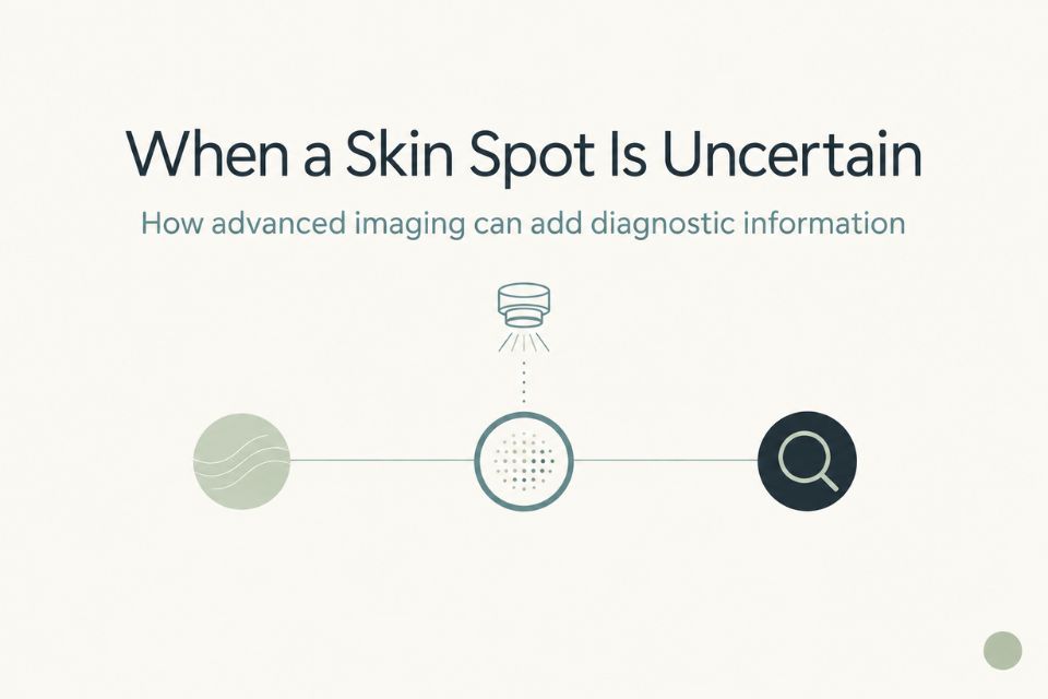
Dermoscopy can provide additional information beyond naked-eye examination. It uses magnification and specialised light to reveal surface and pigment structures that may assist the clinician’s assessment.
A biopsy may be required when the diagnosis is uncertain, invasive SCC is suspected or a lesion is enlarging, thickening, persistently tender, ulcerated, bleeding or not responding as expected.¹˒⁴
Not every rough spot requires biopsy. The decision is based on the individual lesion and the clinical context.
What is the role of dermoscopy and advanced imaging?
Dermoscopy helps clinicians examine lesions in more detail than the naked eye allows. It can support assessment of an AK, help identify features that warrant closer attention and assist in distinguishing lesions that may need biopsy.⁶
Advanced imaging is not required for the routine diagnosis of most straightforward actinic keratoses. However, selected lesions can remain diagnostically uncertain after clinical examination and dermoscopy.
In those cases, reflectance confocal microscopy—also called RCM or optical biopsy—may provide additional non-invasive diagnostic information. RCM allows selected superficial lesions to be viewed at approximately cellular-level resolution without removing tissue.⁷
RCM is not needed for every AK and does not replace surgical biopsy when tissue diagnosis is clinically required. It is best understood as a selective diagnostic tool for an uncertain lesion, rather than a routine test for sun-damaged skin.
Treatment: an individual decision
Skintel is a diagnostic skin imaging service, so treatment planning should be discussed with your treating clinician.
Depending on the number, location and characteristics of AKs, treatment may include cryotherapy, topical or field-directed treatments, photodynamic therapy or other clinician-directed options.²˒⁵
“Field treatment” refers to treatment of a broader area of sun-damaged skin, rather than treating only one visible spot. This may be considered when there are multiple AKs or broader field change.
The appropriate approach depends on the lesion, the extent of sun damage, your general health, previous treatment, likely side effects and personal preferences. Do not start or repeat treatment for a persistent or changing lesion without clinician advice, because a lesion thought to be an AK may require reassessment.
Sun damage and prevention
Actinic keratoses are a reminder that ongoing sun protection matters. Sun protection cannot undo all existing UV damage, but reducing further exposure can help reduce additional damage and support long-term skin health.²˒⁵
For Australian conditions, practical steps include:
-
wearing sun-protective clothing;
-
using a broad-brimmed hat;
-
seeking shade when UV levels are high;
-
wearing sunglasses;
-
applying broad-spectrum SPF 50+ sunscreen to exposed skin; and
-
avoiding unnecessary UV exposure, including deliberate tanning.⁵
Sunscreen is one useful part of sun protection, but it works best alongside clothing, hats, shade and planning outdoor activities around UV exposure.
When should you get an actinic keratosis checked?
Arrange medical assessment if you have:
-
a rough or scaly patch that persists;
-
a lesion that is new or changing;
-
a spot that becomes thicker, firmer or more raised;
-
increasing tenderness or pain;
-
bleeding, ulceration or recurrent crusting;
-
a lesion that does not settle or behaves differently from other rough patches;
-
multiple areas of significant sun damage; or
-
uncertainty about whether a spot is an AK or something else.¹˒⁴
You do not need to treat every stable rough patch as an emergency. However, a changing or concerning lesion should not be ignored, particularly if it is persistent, thickening, tender, bleeding or ulcerated.
How Skintel fits into sun-damage assessment
A comprehensive skin assessment can help identify actinic keratoses, broader signs of cumulative sun damage and lesions that require closer review.
At Skintel, our skin checks include full-body scanning or body mapping, high-magnification assessment of selected lesions and accredited skin cancer doctor review. A detailed report with follow-up recommendations is provided after assessment.⁸
For selected lesions, dermoscopy and other non-invasive imaging may help provide further diagnostic information. If a lesion needs biopsy or treatment, that management is arranged through the patient’s treating or referring clinician. Skintel is a diagnostic skin imaging service and does not provide treatment for actinic keratoses.
Concerned about sun-damaged skin?
If you have persistent rough or scaly lesions, substantial sun damage or another concerning skin spot, a comprehensive skin assessment can help identify areas that need closer clinical attention.
To arrange an assessment, Book a skin check at Skintel.
Medical disclaimer
This article provides general information only. Photographs and descriptions cannot diagnose an actinic keratosis or exclude skin cancer. Persistent, changing, bleeding, painful, thickening, ulcerated or otherwise concerning lesions should be assessed by a qualified medical practitioner. Biopsy may be required when squamous cell carcinoma or another skin cancer is suspected.
Frequently Asked Questions
Is actinic keratosis skin cancer?
No. Actinic keratosis is not the same as invasive skin cancer. It is a sun-damaged lesion with abnormal skin cells that may, in some cases, progress to cutaneous squamous cell carcinoma.¹˒³ Most individual AKs do not inevitably become invasive SCC, but their presence indicates cumulative UV damage and a higher-risk environment for additional keratinocyte cancers.
Can actinic keratosis turn into squamous cell carcinoma?
Yes, some AKs can progress to SCC, but most individual lesions do not inevitably do so.¹˒³ The risk for an individual lesion is difficult to predict. A spot that becomes thicker, rapidly enlarges, tender, firm, ulcerated or prone to spontaneous bleeding should be reassessed because these changes may warrant closer examination or biopsy.
What does actinic keratosis look like?
Actinic keratosis often looks like a rough, scaly, crusted or dry patch on sun-exposed skin. It may be pink, red, brown, pale or skin-coloured.¹˒² Some AKs are easier to feel than see and can have a sandpaper-like texture. These appearances are not diagnostic by themselves, so a persistent or changing rough patch should be assessed by a qualified clinician.
What does actinic keratosis feel like?
Actinic keratosis often feels rough, dry or sandpaper-like.²˒⁴ Some spots are mildly tender or sensitive, while others cause no sensation at all. A rough area that repeatedly returns after scale is removed or remains in the same place for months should be checked, especially if it becomes thicker, painful, bleeding, ulcerated or otherwise changes.
Can actinic keratosis disappear on its own?
Some AKs may regress or appear to disappear, but they can also persist or recur.³˒⁴ A visible rough patch settling does not necessarily mean the surrounding sun damage has resolved. Because it is difficult to predict behaviour and distinguish AK from other lesions by appearance alone, follow your treating clinician’s advice about assessment, treatment and review.
Should actinic keratosis be removed?
Treatment is often recommended, but the right approach depends on the number of lesions, location, severity of sun damage, your medical history and the clinical appearance of the spot.²˒⁵ Options may include lesion-directed treatment, such as cryotherapy, or field treatment for broader sun-damaged areas. Your treating clinician can advise what is appropriate; Skintel provides diagnostic imaging rather than AK treatment.
When should an actinic keratosis be biopsied?
Biopsy may be needed when the diagnosis is uncertain or there are features that raise concern for invasive SCC, such as enlargement, thickening, marked tenderness, firmness, ulceration, spontaneous bleeding or lack of expected response to treatment.¹˒⁴ Not every AK requires biopsy. The decision is based on the lesion’s appearance, behaviour and clinical context.
What is the difference between actinic keratosis and SCC?
An AK is generally a superficial UV-damaged lesion, whereas SCC is an invasive skin cancer that can extend deeper into the skin.¹˒⁴ They can look similar, particularly when both are rough, scaly or crusted. Patients cannot always tell the difference by sight. Clinical examination, dermoscopy and sometimes biopsy are used to clarify the diagnosis.
Does actinic keratosis mean I have severe sun damage?
An AK indicates cumulative UV damage in that area of skin. One lesion does not necessarily mean severe widespread damage, but multiple AKs or rough patches across the face, scalp, ears or hands can indicate broader field change.²˒³ This is a useful reason to review sun protection and discuss an appropriate skin surveillance plan with your clinician.
Can younger people develop actinic keratosis?
Yes. AKs are more common with increasing age because UV damage accumulates over time, but younger people can develop them, particularly with substantial UV exposure, fair or easily sunburned skin or immune suppression.²˒⁵ A persistent rough or changing lesion deserves assessment regardless of age.
Conclusion
Actinic keratosis, also called solar keratosis, is an early visible sign of cumulative sun damage. It commonly appears as a persistent rough or scaly patch on chronically sun-exposed skin and may be easier to feel than see.
AK is not the same as invasive skin cancer, and most individual AKs do not inevitably become SCC. But their presence matters because they indicate UV damage and can be associated with increased future risk of keratinocyte cancer. If a lesion is persistent, changing, thickening, painful, bleeding, ulcerated or simply concerning, arrange medical assessment rather than trying to diagnose it yourself.
References
-
Cancer Council Australia. Clinical practice guidelines for keratinocyte cancer. Sydney: Cancer Council Australia; 2019.
-
O’Bryen J, Green S, Seine AJ, Hudson J, De Cruz R, Reiche A, et al. Pre- and post-treatment care for actinic keratoses: an Australian and New Zealand perspective. Aust J Gen Pract. 2025;54(8).
-
Werner RN, Stockfleth E, Connolly SM, et al. Evidence- and consensus-based (S3) guidelines for the treatment of actinic keratosis: international league of dermatological societies in cooperation with the European Dermatology Forum. J Eur Acad Dermatol Venereol. 2015;29(1):e1-e66.
-
Dodds A, Chia A, Shumack S. Actinic keratosis: rationale and management. Dermatol Ther. 2014;4(1):11-24.
-
Cancer Council Australia. Sun protection: reduce your skin cancer risk. Sydney: Cancer Council Australia; accessed 2026 Sep 7.
-
Kittler H, Pehamberger H, Wolff K, Binder M. Diagnostic accuracy of dermoscopy. Lancet Oncol. 2002;3(3):159-165.
-
Dinnes J, Bamber J, Chuchu N, et al. Reflectance confocal microscopy for diagnosing cutaneous melanoma in adults. Cochrane Database Syst Rev. 2018;12(12):CD013190.
-
Skintel. The Skintel skin check. Melbourne: Skintel; accessed 2026 Sep 7.