
By Dr Maxton Bergin
Skin cancer diagnosis in Australia is changing. Clinical examination and biopsy remain central to care, but doctors can now use more detailed imaging tools than ever before. Dermoscopy, digital photography, mole mapping, total body photography and reflectance confocal microscopy can each add different information about a lesion or a person’s broader skin cancer risk.¹⁻³
Artificial intelligence, or AI, is also beginning to influence this field. AI systems can analyse digital skin images, compare photographs over time and potentially help doctors prioritise lesions for closer assessment. However, AI is not a substitute for a skin cancer doctor, a dermatologist or a biopsy when tissue diagnosis is needed.⁴⁻⁶
The future of skin cancer detection in Australia is likely to involve combining technologies, not replacing one test with another. A patient may have total body photography to identify new or changing lesions, dermoscopy to examine a particular mole, AI-assisted image analysis where clinically validated, reflectance confocal microscopy for a lesion that remains uncertain, and a biopsy when the doctor needs definitive tissue information.
Why advanced skin imaging matters in Australia
Australia has one of the world’s highest burdens of skin cancer. Melanoma is a major health concern, while keratinocyte cancers—including basal cell carcinoma and squamous cell carcinoma—are also extremely common.⁷˒⁸ Early diagnosis is important because many skin cancers can be treated more simply and effectively when found early.⁷˒⁸
At the same time, early detection is not simply about removing every mole. Many Australians have numerous benign moles, freckles and sun-damaged spots. The challenge is to identify lesions that genuinely require biopsy or treatment while avoiding unnecessary procedures where high-quality assessment is reassuring.¹˒²˒⁹
Advanced skin imaging may help clinicians make these decisions more consistently by recording lesions, comparing them over time and adding detail beyond what can be seen with the naked eye.
Technology already used in clinical care
Several advanced imaging methods are already used in Australian skin cancer assessment. Their availability varies between clinics, cities and regional areas.
Dermoscopy
Dermoscopy is a close-up examination of the skin using magnification and specialised polarised light. It helps doctors see pigment patterns, blood vessels and structures beneath the surface that are not visible to the naked eye.¹⁰˒¹¹
Dermoscopy is widely used in skin cancer practice and improves diagnostic accuracy when performed by trained clinicians.¹⁰˒¹¹ It remains the foundation of detailed mole assessment, even as newer technologies develop.
Digital dermoscopy and sequential monitoring
Digital dermoscopic photography records close-up images of individual lesions. At future appointments, the same lesion can be photographed again and compared with earlier images.¹˒²
This is especially helpful for patients with many moles, atypical moles or a higher risk of melanoma. Rather than relying entirely on memory, doctors can examine objective changes in a lesion’s structure, colour or pattern over time.¹˒²
A changing lesion does not automatically mean cancer, but change can be an important reason for further assessment.
Total body photography and mole mapping
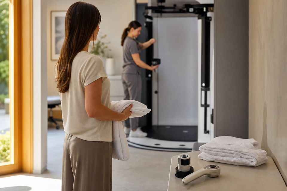
Total body photography creates a photographic record of the skin surface. Mole mapping combines these wider photographs with close-up dermoscopic images of selected lesions.¹˒²
The main benefit is comparison. At later reviews, doctors can look for new lesions or changes in existing lesions. This can be particularly useful for people with many moles or an increased melanoma risk.¹˒²
Reflectance confocal microscopy
Reflectance confocal microscopy, or RCM, is a non-invasive imaging technique that uses a low-power laser to create highly magnified black-and-white images of living skin. It is sometimes called an optical biopsy because it shows cellular-level structures without immediately removing tissue.³˒¹²
RCM is usually used when a lesion remains uncertain after clinical examination and dermoscopy. It is especially helpful in selected facial lesions, sun-damaged skin and cosmetically sensitive areas where biopsy may leave a visible scar.³˒¹²
RCM does not replace biopsy. It does not provide all the information that comes from histopathology—the laboratory examination of tissue removed during a biopsy. If a lesion remains suspicious or uncertain, biopsy may still be necessary.³˒¹²˒¹³
What AI means in skin imaging
In skin imaging, AI usually refers to computer systems trained to identify patterns in medical images. These systems may use machine learning or deep learning, which means they learn from large collections of labelled photographs, dermoscopic images or other clinical images.⁴˒⁵
AI may assist in several ways:
-
analysing a dermoscopic image for patterns associated with benign and malignant lesions;
-
comparing total body or dermoscopic images over time;
-
flagging new or changing lesions for clinician review;
-
helping organise large image collections;
-
supporting triage by highlighting lesions that may need earlier assessment.⁴⁻⁶
AI does not “see” a lesion in the same way a clinician does. It recognises patterns from the data it has been trained on. Its output can be influenced by image quality, lighting, camera type, the type of lesion and how closely the patient resembles the populations included in the training data.⁴˒⁵
Can AI detect skin cancer?
AI can analyse skin images and may help identify lesions that warrant closer assessment, but it cannot independently diagnose or exclude skin cancer for an individual patient.⁴⁻⁶
Some studies show that AI algorithms can perform similarly to experienced dermatologists when tested on selected image datasets. A 2024 systematic review and meta-analysis found that AI performance was clinically comparable with expert dermatologists in the included studies, while differences were greater when AI was compared with generalist clinicians.⁴
However, research studies do not always reflect real-world practice. Many algorithms have limited external validation, use carefully selected images and may not include enough examples of darker skin types, uncommon cancers or images taken outside specialist settings.⁵˒⁶
The most useful role for AI is therefore as decision support: an additional source of information that helps a clinician review images, rather than an automated verdict.
Why AI must be used carefully
AI performance depends on data. If an algorithm is trained mainly on images of lighter skin types or common lesion categories, it may work less reliably for people with darker skin, unusual presentations or less common cancers.⁵˒⁶
A recent systematic review found that representation of skin of colour and cutaneous squamous cell carcinoma was limited in many AI studies, and that external validation was often lacking.⁵ This matters because a system can appear accurate in a research dataset but perform differently in routine clinical care.
False positives and false negatives are also important:
-
A false positive occurs when AI flags a harmless lesion as concerning. This may create anxiety or lead to unnecessary assessment.
-
A false negative occurs when AI does not flag a lesion that is actually cancerous. This could delay diagnosis.
For these reasons, consumer smartphone apps should not be used to diagnose melanoma or to reassure yourself that a changing mole is harmless. Any lesion that is new, changing, bleeding, itchy, painful or otherwise concerning should be assessed by a qualified clinician.⁷˒⁸
The future of mole mapping and total body photography
Digital total body photography is becoming more sophisticated. Newer systems may capture the skin surface quickly, produce three-dimensional body models and support detailed comparison between visits.¹˒²˒¹⁴
In Australia, research programs are investigating whether 3D total body photography combined with computer-assisted analysis can help detect new or changing lesions more efficiently, particularly for people at higher risk of melanoma.¹⁴˒¹⁵
This is promising, but it is still important to distinguish research from routine care. Three-dimensional imaging and AI-assisted comparison are developing rapidly, but their benefits, cost-effectiveness and best use in broad clinical practice continue to be studied.¹⁴˒¹⁶
In the future, these tools may help clinicians focus attention on the lesions most likely to need closer review. They are unlikely to replace the need for a complete clinical examination, particularly in people at higher risk.
How AI, dermoscopy and confocal microscopy may work together
The future of skin imaging may involve a layered diagnostic pathway rather than one “best” test.
A possible pathway could be:
-
Total body photography documents the overall skin surface.
-
Automated image comparison may identify new or changing areas for review.
-
Clinical examination and dermoscopy assess those lesions in person.
-
AI-assisted analysis, where appropriately validated and used, may provide an additional pattern-based opinion.
-
Reflectance confocal microscopy may be used for selected lesions that remain diagnostically uncertain.
-
Surgical biopsy is performed when tissue diagnosis is required.¹⁻⁶˒¹²˒¹³
Not every patient needs every step. A single changing lesion may need immediate biopsy. A person with many moles may benefit from photography and dermoscopic follow-up. A flat facial lesion with uncertain dermoscopic features may benefit from RCM before deciding whether and where to biopsy.
The key point is that these technologies can complement each other. Each has strengths and limitations, and clinical judgement determines which tool is appropriate.
Can improved imaging reduce unnecessary biopsies?
In selected equivocal lesions, high-quality imaging may help clinicians distinguish lesions that need biopsy from lesions that can reasonably undergo further non-invasive assessment or planned monitoring.³˒⁹˒¹²
A randomised clinical trial found that adding RCM to standard assessment reduced the number of lesions excised for each melanoma detected.⁹ This does not mean clinicians should aim for fewer biopsies in general. It means that improved diagnostic specificity may reduce removal of some benign lesions while maintaining a strong focus on detecting melanoma.
This is particularly relevant on the face, nose, eyelids, lips and ears, where an unnecessary biopsy may leave a visible scar or affect delicate anatomy.³˒¹²
What remains experimental or under investigation?
Several emerging technologies may influence future care, but they are not all routine clinical practice.
AI-assisted risk prioritisation
Systems that combine total body photography, dermoscopic images and patient risk factors may eventually help clinicians prioritise lesions for review. This remains under investigation and requires rigorous validation across different settings and skin types.⁴⁻⁶˒¹⁴
Three-dimensional total body imaging
Three-dimensional imaging can create a detailed surface record and may improve lesion tracking. It is available in some specialist and research settings, but its role, cost-effectiveness and ideal patient groups are still being evaluated.¹⁴˒¹⁶
Remote image review and teledermatology
Teledermatology can improve access to specialist advice by allowing clinical or dermoscopic images to be reviewed remotely. This may be particularly valuable in regional, rural and remote parts of Australia.¹⁷
However, remote assessment depends on good image quality, clear clinical information, secure systems and appropriate pathways for urgent in-person review. It cannot replace examination in every situation.¹⁷
AI-assisted confocal microscopy
Researchers are also investigating AI systems that may help organise and analyse RCM images. These systems may assist with image quality, lesion mapping and pattern recognition, but they remain emerging tools rather than routine stand-alone diagnostics.⁶˒¹⁸
Challenges for the future of skin imaging in Australia
Technology can improve access to information, but it also introduces practical and ethical challenges.
Access and cost
Specialised equipment is expensive and requires trained staff. Access may be easier in metropolitan centres than in regional, rural or remote communities.¹⁴˒¹⁷ Expanding access will require workable referral pathways, clinician training and evidence that new tools improve outcomes as well as efficiency.
Clinician training
Dermoscopy, total body photography and RCM are most useful when images are captured and interpreted well. Technology cannot compensate for poor image quality or inadequate clinical assessment.³˒¹⁰˒¹²
Privacy and data security
Skin imaging often involves identifiable photographs, including total body images. Patients need confidence that images are stored securely, used appropriately and handled according to Australian privacy requirements.¹⁹
Regulation and real-world validation
AI tools used in healthcare need careful regulatory oversight, transparent performance testing and ongoing monitoring. Results from research datasets should not automatically be assumed to apply to every clinical setting or patient group.⁴⁻⁶˒²⁰
The continuing role of biopsy
No matter how advanced imaging becomes, biopsy and histopathology will remain essential when a lesion is suspicious, imaging is inconclusive, or tissue information is needed for diagnosis and treatment planning.³˒¹³
Skintel’s role in advanced skin imaging
Skintel is a specialist diagnostic skin imaging service. Its role is to provide advanced diagnostic information that helps referring clinicians make informed decisions about skin lesions and skin cancer risk.
Depending on the patient and referral question, the Skintel diagnostic pathway may include total body photography, mole mapping, digital dermoscopy, computer-assisted image comparison and reflectance confocal microscopy. These findings are reviewed by an experienced skin cancer doctor and documented in a report for the referring clinician.
Skintel does not replace your treating doctor. Your GP, dermatologist, surgeon or other treating clinician remains responsible for ongoing management, biopsy, treatment and follow-up decisions.
Frequently Asked Questions
Can AI detect skin cancer?
AI can analyse skin images and help flag lesions that may need closer review, but it cannot independently diagnose or rule out skin cancer.⁴⁻⁶ AI performance depends on the images and data used to train it, and it cannot reliably assess symptoms, patient history or the full clinical context. A qualified clinician should review concerning lesions and decide whether dermoscopy, imaging or biopsy is required.
Is AI better than a dermatologist at detecting melanoma?
AI has performed similarly to experienced dermatologists in some carefully controlled research studies, but that does not mean it is better in routine care.⁴⁻⁶ Real-world diagnosis involves examining the patient, considering medical history, assessing many lesions and deciding on follow-up or biopsy. Evidence suggests that clinicians may benefit when AI is used as support, rather than as a replacement for clinical expertise.⁶
Can AI diagnose melanoma from a photograph?
No. A photograph alone cannot reliably diagnose melanoma for an individual person. AI systems may analyse an image and flag concerning patterns, but image quality, lighting, skin tone, lesion type and missing clinical information can affect results.⁴˒⁵ A changing or concerning mole should be assessed in person by a qualified clinician.
What is the future of skin cancer detection?
The future of skin cancer detection is likely to combine clinical expertise with digital photography, dermoscopy, computer-assisted comparison, AI-supported analysis and selected high-resolution imaging such as confocal microscopy.¹⁻⁶˒¹² These tools may help clinicians identify change earlier and decide which lesions need biopsy, but biopsy and histopathology will remain essential when tissue diagnosis is required.³˒¹³
Can skin cancer be diagnosed without a biopsy?
Sometimes imaging can provide enough information for a doctor to recommend monitoring rather than immediate biopsy, especially for selected equivocal lesions.³˒⁹˒¹² However, skin cancer cannot always be confirmed or excluded without tissue. Biopsy and histopathology remain necessary when a lesion is suspicious, changing, thick, difficult to image or still uncertain after non-invasive assessment.³˒¹³
What is an optical biopsy?
An optical biopsy is a non-invasive examination, usually reflectance confocal microscopy, that shows highly magnified images of cells and structures in the upper layers of living skin.³˒¹² It does not involve needles or cutting and can be useful for selected lesions that remain uncertain after dermoscopy. It does not replace biopsy when tissue diagnosis is clinically required.
Is confocal microscopy available in Australia?
Yes, reflectance confocal microscopy is available through some specialist skin imaging and dermatology services in Australia, although access is more limited than standard dermoscopy and varies by location.³˒¹² It is usually used for selected lesions rather than as a routine test for every mole. Your doctor can advise whether referral for confocal microscopy is appropriate.
Can mole mapping automatically detect changing moles?
Mole mapping and total body photography can document lesions and help clinicians compare images over time.¹˒² Some systems can assist with identifying new or changing areas, but the images still need clinical review. Automated change detection can be useful, but it cannot determine on its own whether a changing spot is cancerous.¹⁴˒¹⁶
Can a smartphone app diagnose skin cancer?
No. Smartphone apps should not be relied on to diagnose skin cancer or reassure you that a suspicious mole is harmless. AI-based apps may be affected by image quality, lighting, skin tone and limited clinical context.⁴˒⁵ If a spot is new, changing, bleeding, itchy, painful or concerning, arrange a professional skin assessment.
Conclusion
The future of skin cancer detection in Australia is likely to be more connected, data-informed and personalised. Dermoscopy, digital monitoring, mole mapping, total body photography, AI-assisted analysis and reflectance confocal microscopy can each contribute different information to the diagnostic process.
The most important development is not a single machine or algorithm. It is the thoughtful combination of technology with trained clinical judgement. AI may help clinicians organise images, detect change and prioritise lesions, while confocal microscopy may offer additional detail for selected uncertain lesions. But neither replaces biopsy when tissue diagnosis is needed.
For patients, the practical message is simple: advanced technology can support better assessment, but the safest pathway remains one led by experienced clinicians who use the right tool for the right lesion at the right time.
Disclaimer
All information is general and not intended as a substitute for professional advice.
References
-
Tzellos T, Kyrgidis A, Zouboulis CC. The value of total body photography for the early detection of melanoma: a systematic review. J Eur Acad Dermatol Venereol. 2021;35(2):230-237.
-
Salerni G, Carrera C, Lovatto L, et al. Benefits of total body photography and digital dermoscopy monitoring in the early detection of melanoma. J Am Acad Dermatol. 2012;67(1):e17-e27.
-
Longo C, Ragazzi M, Rajadhyaksha M, et al. Reflectance confocal microscopy: an effective tool for diagnosing melanoma and non-melanoma skin cancers. J Am Acad Dermatol. 2018;79(2):247-257.
-
Salinas MP, et al. A systematic review and meta-analysis of artificial intelligence for skin cancer classification compared with clinicians. NPJ Digit Med. 2024;7:119.
-
Hsu DY, et al. Artificial intelligence in dermatology: a systematic review of diagnostic performance, external validation and equity. J Am Acad Dermatol. 2024;91(3):456-467.
-
Tschandl P, et al. Human-AI interaction in skin cancer diagnosis: a systematic review and meta-analysis. NPJ Digit Med. 2024;7:84.
-
Cancer Council Australia. Skin cancer. Sydney: Cancer Council Australia; 2026.
-
Australian Institute of Health and Welfare. Cancer data in Australia: melanoma of the skin. Canberra: AIHW; 2025.
-
Pellacani G, Pepe P, Casari A, Longo C. Reflectance confocal microscopy as a second-level examination in skin oncology improves diagnostic accuracy and saves unnecessary excisions: a longitudinal prospective study. Br J Dermatol. 2014;171(1):104-109.
-
Kittler H, Pehamberger H, Wolff K, Binder M. Diagnostic accuracy of dermoscopy. Lancet Oncol. 2002;3(3):159-165.
-
Menzies SW, Emery J, Staples M, et al. Impact of dermoscopy on the clinical diagnosis of melanoma in primary care: results of a randomised trial. BMJ. 2009;339:b3112.
-
Guitera P, Menzies SW. State of the art of reflectance confocal microscopy for melanoma diagnosis. J Am Acad Dermatol. 2020;83(6):1535-1545.
-
Cancer Council Australia. Clinical practice guidelines for the diagnosis and management of melanoma. Sydney: Cancer Council Australia; 2024.
-
Australian Centre of Excellence in Melanoma Imaging and Diagnosis. 3D total body photography and AI-enabled melanoma screening. Sydney: University of Sydney; 2026.
-
University of Queensland. Tech trial to speed regional melanoma diagnosis. Brisbane: University of Queensland; 2024.
-
Janda M, et al. 3D total body photography with sequential digital dermoscopy imaging for melanoma detection in high-risk patients: randomised clinical trial. JAMA Dermatol. 2025;161(3):245-253.
-
Chan J, et al. Teledermatology for skin cancer assessment in regional and rural Australia: systematic review. Aust J Rural Health. 2022;30(4):445-454.
-
Malciu AM, et al. Artificial intelligence-based approaches to reflectance confocal microscopy in dermatology. Cancers (Basel). 2022;14(2):297.
-
Office of the Australian Information Commissioner. Australian Privacy Principles guidelines. Canberra: OAIC; 2025.
-
Therapeutic Goods Administration. Software-based medical devices: regulation of clinical decision support software. Canberra: Australian Government Department of Health and Aged Care; 2025.