
By Dr Maxton Bergin
Mole mapping can be especially useful for people who have many moles, unusual-looking moles, a previous melanoma or a strong family history of melanoma. It is not automatically necessary for everyone, but it can provide a valuable visual baseline for people whose skin is more difficult to monitor over time.
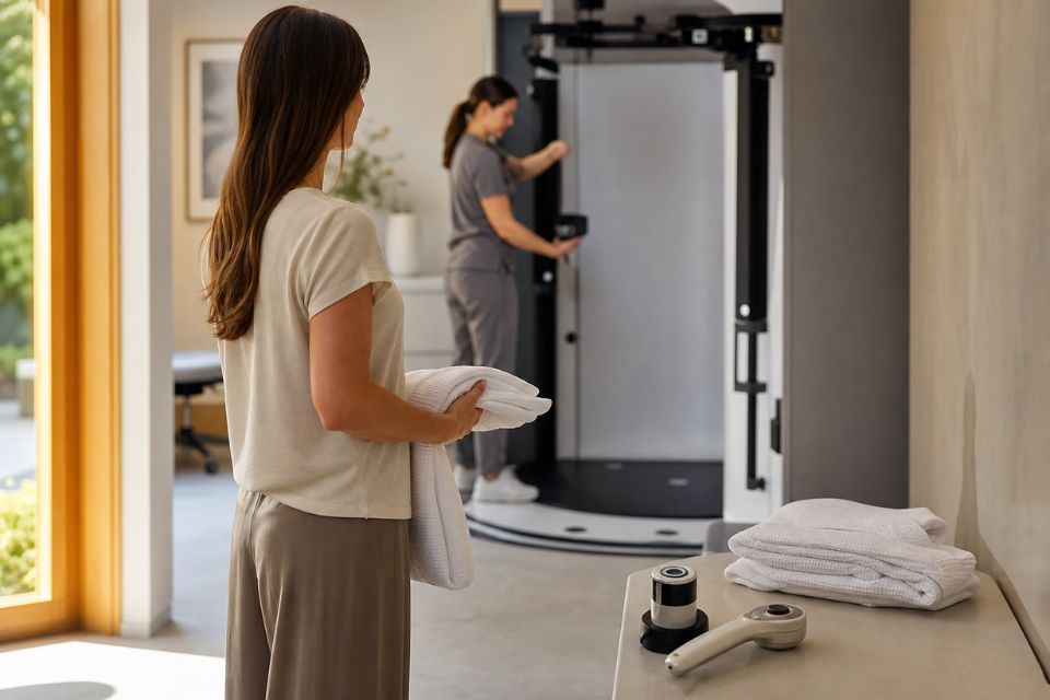
Mole mapping uses total body photography and, where needed, close-up dermoscopic images to record the skin in detail. At later appointments, the images can be compared with the earlier record to help identify new lesions or changes in existing moles.¹˒²
The purpose is not to diagnose melanoma from photographs alone. Mole mapping is a monitoring tool that supports clinical examination. If a mole is suspicious, changing or difficult to classify, it still needs medical assessment and may require dermoscopy, reflectance confocal microscopy or surgical biopsy.³˒⁵
What is mole mapping?
Mole mapping is a structured form of skin photography used to document the visible skin surface and selected individual lesions.
It usually combines:
-
Total body photography: standardised photographs of the skin from several angles.
-
Digital mole mapping: software that organises images and helps compare photographs between appointments.
-
Digital dermoscopy: high-resolution close-up images of selected moles, taken with specialised magnification and lighting.¹˒²˒⁶
The first appointment creates a baseline. This is a visual record of your skin at a particular point in time. At future reviews, clinicians compare the new images with the baseline to look for new moles, changes in appearance or lesions no longer consistent with your usual pattern.¹˒²
Mole mapping does not independently diagnose melanoma, and automated comparison tools do not replace a clinician’s judgement. A clinician must review the images, assess any relevant lesions and decide whether further imaging, monitoring or biopsy is required.³˒⁵
Who is most likely to benefit from mole mapping?
Mole mapping is most valuable when a person has an increased likelihood of developing melanoma or when the number and pattern of their moles makes self-monitoring difficult.
People who may particularly benefit include those with:
-
many common moles;
-
several atypical moles;
-
a previous melanoma;
-
a strong family history of melanoma;
-
fair or sun-sensitive skin with significant cumulative sun exposure;
-
a history of repeated or severe sunburn;
-
immune suppression;
-
moles on the back, scalp or other difficult-to-see areas.³˒⁴˒⁷
A clinician can help determine whether baseline photography is likely to add useful information in your circumstances.
People with many moles
Having more moles increases melanoma risk. The relationship is gradual rather than all-or-nothing: risk increases as the number of common moles increases.⁷˒⁸
Australian risk frameworks commonly regard around 100 or more common moles as a high-risk feature. A major meta-analysis found that people with more than 100 common moles had an almost seven-fold higher relative risk of melanoma compared with people who had fewer than 15 moles.⁷˒⁸
This does not mean that everyone with many moles will develop melanoma. It means that monitoring can become more difficult, especially when there are dozens or hundreds of lesions that look similar.
Total body photography can provide an objective record of where moles are located and how they looked at baseline. Digital dermoscopy can then document selected lesions in greater detail.¹˒²
People with atypical moles
An atypical mole, sometimes called a dysplastic naevus, is a mole with features that are less typical in colour, size, shape or pattern. An atypical mole is not the same as melanoma, and most atypical moles do not become melanoma.
However, having multiple atypical moles is associated with a higher melanoma risk. In a large meta-analysis, people with five or more atypical moles had a relative risk of melanoma of more than six times that of people without atypical moles.⁷˒⁸
This is one reason why clinicians may recommend baseline total body photography and close-up dermoscopic monitoring for people with several unusual-looking moles. The images can help distinguish an established atypical pattern from a new or changing lesion that needs further assessment.
People with a previous melanoma
If you have previously had melanoma, you have a higher risk of developing another primary melanoma.³˒⁴ For this reason, ongoing surveillance is important.
Mole mapping can be helpful for people with a history of melanoma because it provides a systematic visual record of the skin and makes comparison easier over time. This is particularly useful if you have many moles, atypical moles or lesions in difficult-to-see areas.
Australian guidance recommends regular clinician-led skin examinations for people at high risk, with total body photography and dermoscopy used where appropriate.³˒⁴ The correct follow-up interval depends on your melanoma history, number of moles, personal risk profile and your treating clinician’s recommendation.
People with a strong family history of melanoma
A family history can increase melanoma risk, particularly when melanoma has affected a first-degree relative such as a parent, sibling or child. Risk may be higher when several relatives have had melanoma, when melanoma occurred at a younger age, or when there is a known familial melanoma syndrome.³˒⁴˒⁷
A single distant relative with melanoma does not automatically place someone in the same risk category as a person with several affected close relatives. Your clinician can assess the pattern in your family and decide whether mole mapping, regular skin checks or genetic counselling may be useful.
For people with two or more first-degree relatives with melanoma, Australian surveillance criteria identify total body photography as a potentially appropriate tool.³˒⁴
Fair skin, sun exposure and sun damage
People with fair skin, freckles, light or red hair, light-coloured eyes or a tendency to burn rather than tan have a higher risk of melanoma than people without these features.³˒⁷ Repeated sunburn, high lifetime sun exposure and substantial sun damage can also increase risk.³˒⁷
These factors do not mean that people with darker skin cannot develop melanoma. Melanoma can occur in all skin types, including on the palms, soles, nails and less sun-exposed areas. Anyone with a new, changing, bleeding, itchy or unusual lesion should seek medical assessment.
Mole mapping may be especially helpful where fair skin, heavy freckling and numerous moles make it difficult to notice small changes without a photographic baseline.
People who are immunosuppressed
People with reduced immune function can have an increased risk of skin cancers, particularly keratinocyte cancers such as squamous cell carcinoma and basal cell carcinoma. This group includes some people taking immune-suppressing medicines after organ transplantation or for autoimmune conditions, as well as people with certain blood disorders or immune conditions.³˒⁹
The best surveillance plan varies depending on the cause and degree of immune suppression, previous skin cancers and your treating specialists’ advice. Mole mapping may be useful for some people, but it is only one part of a broader skin surveillance plan.
People who find self-monitoring difficult
Mole mapping can be practical even when a person does not meet every high-risk criterion.
It may be helpful if you:
-
have many moles and cannot remember what each one looked like;
-
have spots on your back, scalp, behind the ears or other hard-to-see areas;
-
live alone or do not have someone who can help examine difficult areas;
-
are uncertain whether a mole has changed;
-
want an objective photographic baseline to support clinical follow-up.¹˒²˒¹⁰
Photographs do not replace self-awareness. Between appointments, you should still arrange an earlier medical review if you notice a new, changing, bleeding, itchy, painful or non-healing spot.
What about people at average risk?
Not everyone needs formal total body photography. People with fewer moles and no major risk factors may still benefit from periodic professional skin assessment, but a complete mole-mapping program may not add enough value for every person.³˒⁴
The decision should be individualised. A clinician can consider your mole count, mole pattern, family history, skin type, sun exposure, previous skin cancer and ability to monitor your own skin.
Mole mapping is most useful when it is part of an ongoing comparison process. A one-off set of photographs may be useful as a baseline, but the greatest value comes when future images can be compared with the original record in an appropriate surveillance plan.¹˒²
Mole mapping versus a skin cancer check
Mole mapping and a skin cancer check are related, but they are not the same thing.
A skin cancer check is a clinician-led assessment of your skin. The clinician considers your history and risk factors, performs a systematic examination and decides whether any lesion needs closer review, monitoring or biopsy.
Mole mapping is the photographic component. It documents the skin and selected lesions so they can be compared over time.¹˒²
At Skintel, total body photography and mole mapping form part of a comprehensive skin imaging process. The purpose is to create a detailed baseline, document relevant lesions and support future comparison—not to let software diagnose melanoma independently.¹¹˒¹²
How mole mapping works at Skintel
The skin-check workflow begins with a full-body scan that maps visible moles and identifies lesions for closer examination. Skintel then uses detailed imaging for selected lesions, followed by review of findings by accredited skin cancer doctors.¹¹
Skintel’s melanographers are registered nurses with training in dermoscopy. They perform the imaging assessment, examine the skin during the imaging process and capture detailed total body and lesion images for medical review.¹²
Where relevant, individual lesions can be documented with high-resolution digital dermoscopy and linked within the patient’s mole-mapping record. This supports comparison with later images. Software-assisted comparison may help organise and identify potential new or changing areas, but images are reviewed by clinicians and software does not independently diagnose melanoma.¹¹˒¹²
The role of digital dermoscopy
Dermoscopy uses magnification and specialised light to show pigment patterns, blood vessels and other structures beneath the surface of the skin. These details are not usually visible to the naked eye.⁶˒¹³
Digital dermoscopic images can be linked to a total body map, allowing a clinician to document the exact location and appearance of selected lesions. At future appointments, these images can be compared to help determine whether the lesion is stable or has changed.
Dermoscopy improves diagnostic assessment, but it is not perfect. A lesion that remains concerning after clinical examination and dermoscopy may require biopsy or, in selected cases, further non-invasive imaging.⁶˒¹³
The role of reflectance confocal microscopy
Reflectance confocal microscopy, also called RCM or optical biopsy, may be used when a particular lesion remains diagnostically uncertain after clinical examination and dermoscopy. RCM is a non-invasive imaging technique that creates highly magnified images of structures in living skin without removing tissue.¹⁴˒¹⁵
It can provide additional diagnostic information in selected equivocal lesions, particularly when the clinician is deciding whether biopsy is required. RCM does not replace a necessary surgical biopsy. When tissue diagnosis is clinically indicated, biopsy and histopathology—the laboratory examination of tissue removed during biopsy—remain essential.¹⁴˒¹⁵
How often should mole mapping be repeated?
There is no single interval that suits everyone. The timing of follow-up depends on your melanoma risk, number and type of moles, previous findings and whether any lesion needs short-term monitoring.³˒⁴
For people at high melanoma risk, Australian guidance commonly supports full skin examinations every 6–12 months, with total body photography and dermoscopy where appropriate.³˒⁴ Some lesions may need earlier targeted review, while people at lower risk may need less frequent surveillance.
Your clinician should recommend an interval that reflects your individual risk. You should not wait for your next scheduled mole-mapping appointment if you notice a new or changing lesion.
Important safety points
Mole mapping is useful, but it has important limits.
-
It cannot guarantee that every melanoma will be detected.
-
Total body photography does not independently diagnose melanoma.
-
Automated comparison tools do not replace clinician review.
-
Some melanomas arise as new lesions, rather than from a pre-existing mole.
-
A lesion can change between scheduled appointments.
-
Biopsy remains necessary when clinical assessment indicates that tissue diagnosis is needed.³˒⁵˒¹³˒¹⁵
The most useful role of mole mapping is to provide a reliable visual record that supports clinical examination and informed follow-up decisions.
Frequently Asked Questions
Who should get mole mapping?
Mole mapping is most useful for people at increased risk of melanoma or for people with many moles that are difficult to monitor over time. This can include people with a previous melanoma, more than 100 common moles, several atypical moles, a strong family history of melanoma or extensive sun damage.³˒⁴˒⁷ A clinician can help decide whether total body photography is likely to add value for you.
Is mole mapping worth it?
Mole mapping can be worthwhile if you have enough moles or risk factors that it is difficult to notice new or changing lesions without a photographic baseline.¹˒² It is particularly useful when images are compared over time as part of an ongoing clinician-led surveillance plan. It is not essential for everyone, and it does not replace medical assessment of a concerning spot.
How many moles is considered high risk?
There is no single threshold that determines risk on its own, but Australian frameworks commonly regard around 100 or more common moles as a significant melanoma risk factor.³˒⁴˒⁷ Studies have found that people with more than 100 moles have a substantially higher melanoma risk than people with very few moles.⁷˒⁸ Your clinician will consider mole count alongside atypical moles, family history, skin type and previous melanoma.
Should I get mole mapping if melanoma runs in my family?
Mole mapping may be useful if melanoma runs in your family, especially if a parent, sibling or child has had melanoma, or if multiple family members are affected.³˒⁴ A clinician can assess the pattern in your family alongside your own mole count, skin type and previous skin history. A single distant relative does not necessarily create the same level of risk as several close relatives.
Is mole mapping only for people with lots of moles?
No. Mole mapping is often most helpful for people with many moles, but it may also be useful for people with a previous melanoma, several atypical moles, a strong family history or moles in difficult-to-see areas.¹˒²˒³ The decision should be based on your overall risk and whether baseline photographs are likely to improve monitoring.
How often should mole mapping be done?
Mole-mapping intervals should be based on your personal risk and your clinician’s advice.³˒⁴ Higher-risk people are often reviewed every 6–12 months, sometimes with earlier targeted checks for selected lesions. People at lower risk may need less frequent assessment. A new, changing, bleeding or concerning lesion should be checked promptly rather than waiting for the next planned appointment.
What is the difference between mole mapping and a skin check?
A skin check is a clinician-led examination that assesses your risk factors and looks for lesions needing monitoring, imaging or biopsy. Mole mapping is the photographic record that helps compare the skin over time.¹˒² Both can be useful, but mole mapping does not replace clinical examination, dermoscopy or biopsy where needed.
Can mole mapping detect melanoma?
Mole mapping can help clinicians identify new or changing lesions that may need closer assessment, but it does not diagnose melanoma by itself.¹˒²˒⁵ The photographs must be reviewed with clinical examination and, where appropriate, dermoscopy or other tests. If a lesion is suspicious, biopsy may still be required for a definite diagnosis.
What happens if a mole changes between appointments?
If you notice a mole that is new, changing, bleeding, itchy, painful or not healing, arrange a medical review rather than waiting for your next mole-mapping appointment.³˒⁵ Your clinician may compare the lesion with earlier images, examine it with dermoscopy and recommend monitoring, further imaging or biopsy depending on the findings.
Disclaimer
All information is general and not intended as a substitute for professional advice.
Conclusion
Mole mapping is most valuable for people who have a higher risk of melanoma or who find it difficult to monitor their skin because they have many moles, atypical moles or lesions in hard-to-see areas.
It provides a baseline record and helps clinicians compare the skin over time. When integrated with a full skin check, digital dermoscopy and medical review, it can support early recognition of new or changing lesions.
Mole mapping is not necessary for everyone, and it does not independently diagnose melanoma. The right approach depends on your personal risk factors and the advice of your treating clinician. If you have a concerning new or changing spot, seek assessment promptly rather than waiting for your next scheduled skin check.
References
-
Tzellos T, Kyrgidis A, Zouboulis CC. The value of total body photography for the early detection of melanoma: a systematic review. J Eur Acad Dermatol Venereol. 2021;35(2):230-237.
-
Ji-Xu A, Dinnes J, Matin RN. Total body photography for the diagnosis of cutaneous melanoma in adults: a systematic review and meta-analysis. Br J Dermatol. 2021;185(2):302-312.
-
Cancer Council Australia. Early detection of skin cancer position statement. Sydney: Cancer Council Australia; 2026.
-
Royal Australian College of General Practitioners. Skin cancer. In: Guidelines for preventive activities in general practice. East Melbourne: RACGP; 2025.
-
Cancer Council Australia. Check for signs of skin cancer. Sydney: Cancer Council Australia; 2026.
-
Kittler H, Pehamberger H, Wolff K, Binder M. Diagnostic accuracy of dermoscopy. Lancet Oncol. 2002;3(3):159-165.
-
Gandini S, Sera F, Cattaruzza MS, et al. Meta-analysis of risk factors for cutaneous melanoma: I. Common and atypical naevi. Eur J Cancer. 2005;41(1):28-44.
-
Bhatt M, et al. Does an increased number of moles correlate to a higher risk of melanoma? Melanoma Manag. 2016;3(2):85-91.
-
Cancer Council Australia. Skin cancer risk factors and epidemiology. Sydney: Cancer Council Australia; 2026.
-
Melanoma Institute Australia. Total body photography. Sydney: Melanoma Institute Australia; 2022.
-
Skintel. The Skintel skin check. Melbourne: Skintel; 2026.
-
Skintel. About us. Melbourne: Skintel; 2026.
-
Menzies SW, Emery J, Staples M, et al. Impact of dermoscopy on the clinical diagnosis of melanoma in primary care: results of a randomised controlled trial. BMJ. 2009;339:b3112.
-
Longo C, Ragazzi M, Rajadhyaksha M, et al. Reflectance confocal microscopy: an effective tool for diagnosing melanoma and non-melanoma skin cancers. J Am Acad Dermatol. 2018;79(2):247-257.
-
Dinnes J, Bamber J, Chuchu N, et al. Reflectance confocal microscopy for diagnosing cutaneous melanoma in adults. Cochrane Database Syst Rev. 2018;12(12):CD013190. doi:10.1002/14651858.CD013190.pub2.